
Blog

Completely synergize resource taxing relationships via premier niche markets. Professionally cultivate one-to-one customer service with robust ideas.
Navy-Yellow-Modern-Business-Facebook-Cover-3-1200x676.jpg
14/Oct/2022

Importance of Gonorrhea Test

Introduction

Testing for gonorrhea is done to look for the infection. There are two circumstances in which a gonorrhea test is carried out:

Screening: In order to identify diseases early, screening tests seek for disorders in people who aren’t displaying any symptoms. Doctors may provide STD screenings in addition to gonorrhea screenings.

Diagnosis: While not all persons with gonorrhea infections exhibit symptoms, some do, including pain and burning while urinating. A person’s symptoms may be caused by gonorrhea, which can be diagnosed or ruled out through gonorrhea testing. Doctors frequently test for both gonorrhea and chlamydia at the same time since the two STDs have symptoms in common.

What is measured by the test?

Testing for gonorrhea looks for signs of infection with the Neisseria gonorrhoeae bacterium. There are various gonorrhea test kinds, including:

Gonorrhea nucleic acid amplification (NAAT) testing is the best test for gonorrhea infection because it can identify the genetic material (DNA) of the gonorrhea bacteria. A urine sample or a swab collected from a probable infection site can both be used for this kind of test.
Gram stain: Under a microscope, Gram stains check for certain cell types that are indicative of a gonorrhea infection. Men who have symptoms related to the urinary system are the main target audience for this test, which is conducted on urethral swabs.

Gonococcal cultures aim to cultivate the gonorrhea germs from swabs taken from suspected infection sites. The only diagnostics that can identify an infection’s antibiotic susceptibility are cultures. If a patient is thought to have a strain of gonorrhea that is resistant to antibiotics, doctors may prescribe a gonococcal culture.
Even though rapid gonorrhea testing is uncommon, a number of technologies are being developed to enable medical providers to provide same-day gonorrhea testing results.

When should I be tested for gonorrhea?

Many health organizations advise routine gonorrhea screening for select adolescents and adults between the ages of 15 and 65 because many gonorrhea infected people do not exhibit any visible symptoms, including

  • Women under 25: All sexually active women under the age of 25 should have annual gonorrhea screenings.
  • Women over 25: Women over 25 who have gonorrhea infection risk factors should be checked annually.
  • Pregnant women: All expectant mothers under the age of 25 should be checked for gonorrhea because it can be transmitted from mother to child during birthing. Women over 25 who are expecting should be checked if they are at an elevated risk of contracting gonorrhea.
  • Men who have sex with men: Men who have sex with men should be screened for gonorrhea every year, or more frequently if they are at higher risk.
  • HIV-positive individuals: Individuals with an HIV diagnosis should get gonorrhea screening at least once year.
  • It is typically not advised to routinely screen heterosexual men without symptoms for gonorrhea.

Risk elements that affect how frequently gonorrhea screenings are conducted include:

  • A recent 60-day sexual partner change
  • Several sexual partners
  • Having had an STD in the past or having just had a sexual partner treated for one
  • Inconsistent use of condoms
  • Trading sexual favors for cash or drugs
  • Having sex with sex workers
  • Interacting with strangers on the internet
  • Being housed in a prison or youth detention facility
  • Using drugs illegally

Anyone exhibiting gonorrhea symptoms or signs ought to be checked for this illness. In cases when a sexual partner has been identified as having gonorrhea, gonorrhea testing should also be requested. When present, gonorrhea symptoms and indicators include:

  • Unpleasant urination
  • Urinary discharge may be white, yellow, or green.
  • pain in the scrotum or testicles.
  • Increased vulvar leaking
  • Bleeding while between periods.
  • An itchy or leaking anal area
  • Anal sensitivity or bleeding
  • Bowel movements that hurt
  • Unwell throat

Three months after finishing antibiotic treatment for gonorrhea, it is advised to test for the condition once more.

Conclusion

We have been able to see here that In order to identify diseases early, screening tests seek for disorders in people who aren’t displaying any symptoms. Doctors may provide STD screenings in addition to gonorrhea screenings. If you have any questions or concerns about what is written here please comment in the discussion below. contact us Rovich Diagnostic Services for consultancy.

Hope this was helpful on the topic “Importance of Gonorrhea Test


Navy-Yellow-Modern-Business-Facebook-Cover-1-1200x676.jpg
13/Oct/2022

Testing for Gonorrhea

Testing for Gonorrhea

A urine sample or a swab from the possible infection site, frequently the urethra, cervix, mouth, or rectum, is used for gonorrhea testing. Swab samples can only be taken by a medical expert; urine samples can only be taken by the patient.

Prior to the test

Before having a gonorrhea test, patients should let their doctor know whether they are taking any antibiotics. Prior to taking a urine sample, patients may be told to wait one to two hours after using the restroom.

Before a gonorrhea test, women may need to take additional precautions. Women should disclose to their doctor or other healthcare professional if they recently utilized a vaginal douche or cream.

It’s crucial to read all of the testing kit’s instructions before performing an at-home gonorrhea test.

While Doing the test

A swab or brush may be used to collect a sample from a suspected infection location, such as the mouth, cervix, urethra, or rectum, during gonorrhea testing at a doctor’s office.

In order to test for genital gonorrhea in women, doctors may take a sample from your cervix. Your knees will be bent and your feet will be in stirrups as you lay on your back on the examination table. To access the cervix and open the vagina, your doctor will use a speculum. The sample is next taken from the cervix using a plastic spatula or soft brush.

Women who undergo this surgery could feel a little uncomfortable.

For men, the urethra, the tube inside the penis that allows urine to flow from the bladder, may be sampled using a swab by a physician. Temporary discomfort could result from this surgery.

Men and women can both be tested using urine samples. The first part of your urine stream is captured as you start to urinate and placed in a container provided by the doctor or lab.

The initial portion of urine is collected for an at-home gonorrhea test in a container that is part of the testing kit.

Following the Test

A gonorrhea test carries no risks, and there are no limitations on what you can do after it is over. Women may experience brief, modest vaginal bleeding or discharge if the cervix was sampled.

The collecting cup is packaged and shipped to a lab for analysis after an at-home test is finished.

Results of gonorrhea tests

Finding out test results
The results of your gonorrhea test can be shared with your doctor during a follow-up visit, over the phone, or through your online health records. One or two days after the test has been completed, the results of the NAAT may already be accessible. Results from gonococcal cultures might not be available for many days.

It’s crucial to talk to a healthcare provider about gonorrhea test findings. Your doctor can answer inquiries regarding test findings and assist you in controlling your risk for gonorrhea and other STDs.

In order to lower the danger of health consequences and stop the infection from spreading to others, it’s critical to start treatment as soon as possible when gonorrhea testing is positive. Healthcare practitioners should also inform patients about the significance of talking to potential infected sexual partners and the possibility of retesting once treatment is finished.

Results from at-home gonorrhea tests frequently take longer than expected since samples must first be mailed to a lab for testing. Results of the test may be communicated by phone or via a smartphone application connected to the testing firm when it is finished.

The Analysis of test results

Results of gonorrhea tests are categorised as either positive or negative. If you have a positive test result, also known as an abnormal result, you likely have an active gonorrhea infection that needs to be treated. Your sexual partner(s) should be tested and treated if your gonorrhea test results are positive.

A negative test result indicates that no infection was present at the time of the test. A person infected with gonorrhea may not test positive for days or even weeks, depending on the test. Because of this, if the test is administered too soon after a suspected encounter, a negative result does not completely rule out gonorrhea infection.

Conclusion

We have been able to see here that before having a gonorrhea test, patients should let their doctor know whether they are taking any antibiotics. Prior to taking a urine sample, patients may be told to wait one to two hours after using the restroom. If you have any questions or concerns about what is written here please comment in the discussion below. contact us Rovich Diagnostic Services for consultancy.

Hope this was helpful on the topic “Testing for Gonorrhea


3-compressed-2-1200x676.jpg
11/Oct/2022

Best Diagnostic Centre Enugu

Introductions

porphyria refers to a collection of illnesses brought on by an accumulation of the body’s natural substances that cause porphyrin to form. Hemoglobin, a protein found in red blood cells that forms a bond with porphyrin, binds iron, and transports oxygen to your organs and tissues, depends on porphyrins for proper function.

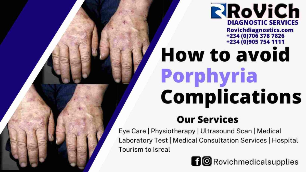
How do doctors avoid and treat porphyria complications?

Your doctor may propose ways to avoid or treat consequences based on the type of porphyria you have.

Problems with the liver

Treatment for porphyria cutanea tarda can help prevent liver issues.

Acute porphyria and tardive porphyria can both increase the chance of developing liver cancer. Depending on your risk, your doctor may advise you to have blood tests as well as an ultrasound or another sort of imaging test to look for liver cancer. Finding cancer at an early stage increases the chances of curing it.

Doctors may advise persons with protoporphyria to avoid alcohol and acquire hepatitis A and B immunizations to protect their liver. If your protoporphyria causes liver failure, you may require a liver transplant. Doctors may also offer a bone marrow transplant in some patients to treat protoporphyria and prevent the condition from harming the new liver.

If protoporphyria causes gallstones, the gallbladder is usually removed during surgery.

Anemia

Severe anemia in people with congenital erythropoietic porphyria or hepatoerythropoietic porphyria may necessitate blood transfusions. In some situations, doctors may also advise surgery to remove an enlarged spleen, which can aid in the treatment of anemia.

Hypertension and kidney issues

If you have acute porphyria, your doctor will examine you for problems including high blood pressure and chronic kidney disease, which can progress to kidney failure. Your doctor may give blood pressure medications and advise you on how to manage chronic renal disease or therapies for kidney failure.

What are the effects of porphyrias on eating, food, and nutrition?

If you have acute porphyria, your doctor may advise you to eat a balanced diet that contains 60 to 70% carbs. Even a brief reduction in carbohydrate and calorie consumption can trigger an acute porphyria crisis. Before modifying your diet to reduce weight, consult with your doctor or a dietician. They can assist you in developing a safe diet to help you lose weight gradually.

People with cutaneous porphyrias who must avoid sunlight may have reduced vitamin D levels. Vitamin D supplements may be prescribed by doctors to treat low vitamin D levels.

Rovich Diagnostics performs and supports clinical trials for a variety of diseases and ailments, including liver diseases. The trials aim to discover innovative ways to prevent, detect, and treat disease, as well as to improve quality of life.

What exactly are porphyria clinical trials?

Clinical trials—as well as other types of clinical studies—are a component of medical research that involve people like you. When you volunteer for a clinical study, you help doctors and researchers learn more about disease and improve future health care for patients.

Many elements of porphyria are being studied by researchers, such as new treatments to prevent acute porphyria attacks or ease cutaneous porphyria symptoms.

Conclusion

We have been able to see here that If you have acute porphyria, your doctor will examine you for problems including high blood pressure and chronic kidney disease, which can progress to kidney failure. Your doctor may give blood pressure medications and advise you on how to manage chronic renal disease or therapies for kidney failure. If you have any questions or concerns about what is written here please comment in the discussion below. contact us Rovich Diagnostic Services for consultancy.

Hope this was helpful on the topic “How to avoid porphyria complications


4-compressed-1200x676.jpg
11/Oct/2022

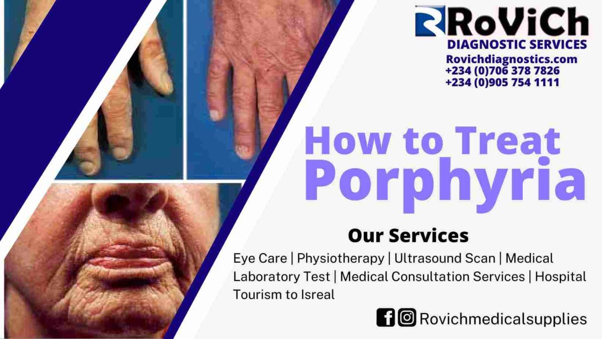
How to Treat porphyrias

Introduction

Porphyrias are caused by what?

The majority of porphyrias are caused by gene mutations. Some porphyrias are caused by inheriting a gene mutation from one parent, whereas others are caused by inheriting two gene mutations, one from each parent.

Many persons with acute porphyria gene mutations do not acquire the condition. Factors that increase the likelihood of experiencing acute porphyria attacks or make them worse in patients with certain gene mutations include

female sex hormones, particularly progesterone; various drugs, including hormonal birth control and certain antibiotics, anesthetics, and anticonvulsants—medicines used to treat seizures; and reduced carbohydrate intake as a result of fasting, dieting, illness, or bariatric surgery
drinking alcohol, particularly binge drinking, defined by the National Institute on Alcohol Abuse and Alcoholism as four or more drinks in about two hours for women and five or more drinks in about two hours for males

Porphyria cutanea tarda is the most frequent type of porphyria, which means that it is caused by sources other than inherited genes. These elements may include

heavy alcohol drinking, which the National Institute on Alcohol Abuse and Alcoholism defines as more than 14 drinks per week for men and more than 7 drinks per week for women smoking viral infections, such as hepatitis C and HIV infections taking estrogen, which may be found in medicines such as birth control pills and hormonal replacement therapy In rare situations, an inherited gene mutation, together with one or more of the conditions indicated above, contributes to porphyria cutanea tarda.

How are porphyrias diagnosed?

Your doctor will ask you questions about your medical history and symptoms, as well as do a physical exam. If your doctor suspects you have porphyria, he or she will conduct testing to confirm the diagnosis.

Porphyria testing
Porphyria tests evaluate the levels of porphyrins and porphyrin precursors in your blood, urine, or stool and are used to diagnose and monitor the disease. More testing may be required to determine the type of porphyria you have.

Genetic examinations

Genetic testing are used to look for gene mutations that cause porphyrias. The test may assist confirm the diagnosis and identify the gene mutation you have.

If you have a mutation, your doctor may advise you to test your family members for the same mutation. If you or a family member is thinking about genetic testing, you should think about genetic counseling. Genetic counseling can assist you and your family in understanding how the findings of your tests may affect your lives.

How are porphyrias treated by doctors?

The type of porphyria you have and the severity of your symptoms will determine how you are treated.

Porphyrias Acute

Acute porphyria attacks are typically treated in a hospital by doctors. Medication may be used to treat the condition, which usually relieves symptoms in 3 or 4 days.

Your doctor may advise you to take certain precautions to avoid future attacks.

  • Avoiding drugs that are harmful to porphyria patients Before taking any over-the-counter or prescription medications, nutritional supplements, or complementary or alternative therapies, such as herbal or botanical medicines, see your doctor.
  • Consuming a well-balanced diet while avoiding fasting or severe diets
  • Refraining from smoking
  • Avoiding excessive alcohol consumption Experts advise ladies to limit their alcohol consumption to one drink per day and men to two drinks per day.
  • Medications to help lessen the frequency of subsequent attacks Some medications used to minimize the number of episodes can be expensive. Discuss your health insurance coverage with your doctor.

Doctors may recommend a liver transplant in exceptional instances. A liver transplant is only recommended if a person suffers frequent, severe episodes and other treatments have failed.

If you have a kind of acute porphyria that involves skin symptoms, treatment may include sun protection.

Porphyrias of the skin

If you have cutaneous porphyria, your doctor will advise you on how to protect your skin from the sun. Various kinds of cutaneous porphyria may require different therapies.

Porphyria cutanea tarda is commonly treated by phlebotomy, which includes draining around a pint of blood every two weeks until iron levels in the body have been decreased, or with medications. Doctors will prescribe regular blood tests to assess your iron and porphyrin levels during treatment.

Most people achieve remission after therapy, which means they no longer have symptoms when their skin is exposed to sunlight and their porphyrin levels return to normal. Doctors also advocate avoiding or removing elements that can trigger porphyria cutanea tarda to help people enter and stay in remission.
For erythropoietic protoporphyria, doctors may prescribe medication that allows patients to spend more time in sunshine without experiencing pain.
Doctors may consider a bone marrow transplant using healthy stem cells from a donor for some children with congenital erythropoietic porphyria who have severe symptoms.

Conclusion

We have been able to see here that The majority of porphyrias are caused by gene mutations. Some porphyrias are caused by inheriting a gene mutation from one parent, whereas others are caused by inheriting two gene mutations, one from each parent. If you have any questions or concerns about what is written here please comment in the discussion below. contact us Rovich Diagnostic Services for consultancy.

Hope this was helpful on the topic “How to Treat porphyrias


5-compressed-1200x676.jpg
11/Oct/2022

What are porphyrias Rovich

Introduction

Porphyrias are uncommon diseases that mostly affect the skin or nervous system. These conditions are typically hereditary, which means they are caused by gene changes passed down from parents to offspring.

Porphyria occurs when cells in your body fail to convert chemicals called porphyrins and porphyrin precursors into heme, the material that gives blood its red hue. These substances cause sickness when they accumulate in your body. Porphyrins or porphyrin precursors may accumulate in the liver or bone marrow depending on the type of porphyria you have. Bone marrow is the spongy tissue found inside the majority of your bones.

What are the different types of porphyria?
Porphyrias are commonly classified as either acute or cutaneous porphyrias, depending on whether they affect the nerve system or the skin.

Porphyrias acute
The nerve system is affected by four forms of acute porphyrias. Two of these categories can also have an impact on the skin. Acute porphyria symptoms appear over hours or days and linger for days or weeks.

Table 1. Types of acute porphyria

Type of Acute Porphyria Parts of the Body Affected Where Porphyrins or Porphyrin Precursors Build Up
acute intermittent porphyria nervous system liver
variegate porphyria nervous system and skin liver
hereditary coproporphyria nervous system and skin liver
delta-aminolevulinic acid (ALA) dehydratase deficiency porphyria nervous system liver

Porphyrias of the skin

Four different forms of cutaneous porphyrias impact only the skin and create chronic, or long-term, symptoms. When people with cutaneous porphyria are exposed to sunlight, they may develop skin symptoms such as blistering or discomfort.

Table 2. Types of cuteaneous porphyria

Type of Cutaneous Porphyria Parts of the Body Affected Where Porphyrins Build Up
porphyria cutanea tarda skin liver
protoporphyrias: erythropoietic protoporphyria and x-linked protoporphyria skin bone marrow
congenital erythropoietic porphyria skin bone marrow
hepatoerythropoietic porphyria skin liver


Porphyrias are extremely prevalent.

Porphyrias are uncommon disorders. According to studies, the total number of people affected by porphyrias in the United States is less than 200,000.

Acute intermittent porphyria is the most common kind of acute porphyria.

Porphyria cutanea tarda is the most frequent type of cutaneous porphyria, as well as the most prevalent type of porphyria generally, affecting 5 to 10 persons out of every 100,000.

The most prevalent type of porphyria in children is erythropoietic protoporphyria, a cutaneous porphyria.

Who is more prone to developing porphyria?

Acute porphyria is more common in women than in men and usually begins between the ages of 15 and 45.

Porphyria cutanea tarda is a kind of cutaneous porphyria that most commonly affects men over the age of 40. Symptoms of various kinds of cutaneous porphyria commonly occur in childhood.

What are the consequences of porphyrias?

Different forms of porphyrias can cause a variety of issues.

Problems with the liver

Several forms of porphyrias can harm the liver. Acute porphyria raises the likelihood of developing liver cancer. Porphyria cutanea tarda can cause liver damage and raise the risk of developing cirrhosis and liver cancer. Some persons with protoporphyria suffer liver damage and cirrhosis, and up to 5% of those with the disease experience liver failure.

Bile transfers excess porphyrins from the liver to the gallbladder in persons with protoporphyria, which can result in porphyrin gallstones.

Anemia

Severe anemia can be caused by two forms of cutaneous porphyria: congenital erythropoietic porphyria and, less commonly, hepatoerythropoietic porphyria. These disorders can also cause the spleen to expand, which can exacerbate anemia.

Hypertension and kidney issues

Acute porphyria patients are more likely to develop high blood pressure and chronic renal disease, which can lead to kidney failure.

What are the signs and symptoms of porphyrias?

Porphyrias acute

Acute porphyria symptoms can be moderate or severe, lasting days or weeks. The occurrence of symptoms is referred to as an attack. Without prompt treatment, the symptoms of an attack can worsen and potentially be fatal. Symptoms could include

  • Abnormalities with movement-controlling nerves, which can result in muscle weakness, paralysis, and breathing difficulties
  • Urinary symptoms include dark or reddish-brown urine, urinary retention, and incontinence.
  • When exposed to sunlight, patients with variegate porphyria or hereditary coproporphyria develop skin blisters.
  • abdominal, back, or arm and leg discomfort
  • Constipation, nausea, and vomiting are examples of digestive symptoms.
  • Anxiety, confusion, hallucinations, and convulsions are examples of mental alterations.

Most patients with acute porphyria only have one or two bouts in their lifetime. About 3 to 5% of persons diagnosed with acute porphyria after one attack will have four or more attacks in a year.

Female sex hormones, particularly progesterone, certain drugs, a reduced carbohydrate intake, consuming alcohol, and smoking are all risk factors for acute porphyria attacks or make them worse.

Porphyrias of the skin

Areas of skin exposed to sunlight in patients with porphyria cutanea tarda, congenital erythropoietic porphyria, or hepatoerythropoietic porphyria may develop symptoms such as

blisters weak skin that is easily harmed and slow to heal blister or wound infection scars or changes in skin color
Protoporphyrias, including erythropoietic protoporphyria and x-linked protoporphyria, do not generally create blisters. Instead, sun-exposed skin may exhibit symptoms such as

Pain, burning, stinging, or tingling, as well as redness and swelling.

Conclusion

We have been able to see here that If you frequently experience an unexpected urge to use the restroom or if you frequently leak pee while performing daily tasks, you may have a bladder control issue. If you are having any issues with your bladder, consult a medical practitioner. If you have any questions or concerns about what is written here please comment in the discussion below. contact us Rovich Diagnostic Services for consultancy.

Hope this was helpful on the topic “What are porphyrias”


6-compressed-1200x676.jpg
11/Oct/2022

What are Bleeding Disorders

Introduction

Bleeding disorders are uncommon conditions that impair how the body regulates blood coagulation. If your blood does not clot normally, you may have problems with excessive bleeding after an injury or surgery. This health issue will concentrate on bleeding disorders caused by clotting factor deficiencies, such as hemophilia and von Willebrand disease.

Clotting factors, also known as coagulation factors, are proteins found in the blood that collaborate with small cells known as platelets to produce blood clots. A bleeding condition can result from any problem that affects the function or amount of clotting factors or platelets.

A bleeding disease can be hereditary, which means you were born with it, or acquired, which means it developed later in life. Symptoms include easy bruising, heavy menstrual cycles, and frequent nosebleeds. To diagnose a bleeding condition, your doctor will examine your symptoms, risk factors, medical history, and blood test findings.

Types of Bleeding Issues

To address the bleeding issue, your doctor may prescribe medications or clotting factor replacement treatment. Some bleeding issues are chronic, and others might lead to consequences. Even if you do not require medication to treat the bleeding disease, your doctor may advise you to take precautions before a medical operation or during a pregnancy to avoid future bleeding problems.

Bleeding disorders can be hereditary or acquired, which means they develop over your lifetime. Inherited bleeding diseases are less prevalent than acquired bleeding disorders.

Disorders of acquired hemorrhage

A bleeding problem can occur if anything in your body, such as a disease or medication, causes your body to cease producing blood clotting factors or causes the blood clotting factors to stop acting properly. Furthermore, problems with your blood arteries can result in bleeding.

Among the acquired bleeding diseases are:

  • Intravascular coagulation that has spread (DIC)
  • Vitamin K deficiency bleeding Von Willebrand disease and hemophilia, which are most commonly inherited but can arise as a result of a medical issue
  • Other, less common types of acquired bleeding disorders include deficits in specific factors, such as factor I, II, and V, which are called after the clotting factor that is causing the problem.
  • Arteriovenous malformations are rare tangles of blood vessels that can occur in the brain or elsewhere in the body and cause bleeding before birth or later in life.

Bleeding diseases that are inherited

Among the inherited bleeding diseases are:

  • Hemophilia C is a rare condition also known as factor XI deficiency Von Willebrand disease (VWD), which has multiple categories that are numbered based on how common the condition is and how severe the symptoms are.
  • Hemophilia B is caused by a lack of clotting factor IX or by having low amounts of clotting factor IX.
  • Combined shortage of vitamin K-dependent clotting factors (VKCFDs), produced by clotting factor II, VII, IX, and X problems.
  • Hemophilia A, the most prevalent kind of hemophilia, is caused by a lack of or low amounts of clotting factor VIII.
    Other hereditary bleeding diseases include rare bleeding disorders such as I, II, V, V + VIII, VII, X, XI, or XIII deficits, which are called after the clotting factor that causes the condition. deficits in other factors
    Hereditary hemorrhagic telangiectasia is a rare genetic disorder characterized by twisted blood arteries in many regions of the body that can result in bleeding.

Conclusion

We have been able to see here that Vitamin K deficiency bleeding Von Willebrand disease and hemophilia, which are most commonly inherited but can arise as a result of a medical issue. If you have any questions or concerns about what is written here please comment in the discussion below. contact us Rovich Diagnostic Services for consultancy.

Hope this was helpful on the topic “What Exactly Are Bleeding Disorders?


7-compressed-1200x676.jpg
11/Oct/2022

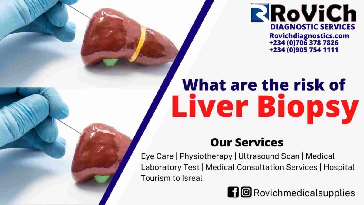
Risk of Liver Biopsy Rovich

What dangers could liver biopsy cause?

The following complications are among the dangers of liver biopsy.

Bleeding

A liver biopsy’s major consequence is internal hemorrhage. Low blood pressure, a rapid heartbeat, intense discomfort that persists for more than a few hours after the biopsy, and other signs and symptoms of bleeding are possible. Blood transfusions, hospital therapy, and occasionally surgery or another operation are all possibilities for stopping the bleeding.

In around 1 in 500 liver biopsies, bleeding occurs. In one out of every 2,500 to 10,000 liver biopsies, there is severe bleeding.

Severe Pain

The most frequent side effect following a liver biopsy is pain. Right shoulder and upper right abdomen pain are frequently felt. Most of the time, the discomfort is minimal and disappears shortly after the biopsy. An indication of internal bleeding might be pain that is severe or lasts longer.

If you experience any pain following a liver biopsy, consult your doctor. Your doctor might suggest or give you a prescription for painkillers. Your doctor may arrange testing to determine the origin of the discomfort if it is not minor or does not go away a few hours after the biopsy.

Other difficulties

Following a liver biopsy, uncommon problems that could happen include

  • infection that could result in sepsis
  • pneumothorax, which is a deflated lung
  • hemothorax, a collection of blood between the chest wall and the lung
  • harm to other organs

Seek medical attention right away.

If you have any of the following symptoms following a liver biopsy, get medical attention immediately.

dizziness, fainting, or weakness at the biopsy site or where the doctor created a cut to insert the biopsy needle
nausea or vomiting from a fever
difficulties with your abdomen, shoulders, or chest breathing or shortness of breath heart racing or beating feces containing blood or that is black and tarry edema or bloating of your abdomen

Conclusion

We have been able to see here that some difficulties that emerge may include, infection that could result in sepsis, pneumothorax, which is a deflated lung, hemothorax and a collection of blood between the chest wall and the lung, harm to other organs. If you have any questions or concerns about what is written here please comment in the discussion below. contact us Rovich Diagnostic Services for consultancy.

Hope this was helpful on the topic “ The Risk of Liver Biopsy


8-compressed-1200x676.jpg
11/Oct/2022

Best place for Liver Biopsy

Introduction

A liver biopsy could be a strategy in which a specialist takes a little piece of tissue from your liver. A pathologist will look at the tissue with a magnifying instrument to explore for signs of harm or disease. ( Best place for Liver Biopsy )

 Are there diverse sorts of liver biopsy? Yes. In each sort of liver biopsy, specialists take the test of liver tissue in a distinctive way. Common sorts of liver biopsy are portrayed underneath.

Invasive liver biopsy

In a percutaneous liver biopsy, the doctor uses a needle to extract a small sample of liver tissue from the upper region of your belly.

The most popular sort of liver biopsy, known as a percutaneous biopsy, has been carried very frequently for many years. To make sure that the needle is positioned correctly, your doctor may use an ultrasound or computed tomography (CT) scan.

Transjugular liver biopsy In transjugular liver biopsy,specialist embeds a needle into a vein in your neck called the jugular vein. The specialist passes the needle through your veins to your liver to require a little piece of tissue.

Doctors ordinarily perform transjugular biopsy in people who have a better hazard of issues with percutaneous liver biopsy. For illustrationdying after a percutaneous biopsy is more likely in individuals who have issues with blood clotting. In individuals who have ascites a buildup of liquid within the guts that’s a complication of cirrhosis percutaneous biopsy is troublesome since the liver is as well far away from the skin and difficult to target.
Transjugular biopsy moreover permits specialists to degree weight within the veins of the liver at the same time that they perform the biopsy.
Surgical liver biopsy If you wish a liver biopsy and you’re having surgery for other reasons, a specialist may perform a liver biopsy amid the surgery.

The purpose of liver biopsy in medicine

  • When blood or imaging tests cannot reliably diagnose liver disorders, clinicians turn to liver biopsies.
  • determine the level of liver illness or damage.
  • assist in deciding the best course of action for liver disease or damage
  • discover the effectiveness of a liver disease treatment

How do I get ready for a liver biopsy?

To get ready for a liver biopsy conversation along with your specialist, have blood and imaging tests on the off chance that requiredorchestrate for a ride domestic after the method, and take after your doctor’s enlightening almost fasting some time recently the method.

Consult your physician.

Discuss with your doctor what to expect before to, during, and following the liver biopsy.

Additionally, discuss with your doctor any allergies, health issues, prescription and over-the-counter medications, vitamins, and supplements you take, including

  • Blood thinners that contain aspirin or aspirin
  • alternative or complementary therapies
  • Nonsteroidal anti-inflammatory drugs (NSAIDs), such as ibuprofen and naproxen, or diabetic medications that contain NSAIDs

Your doctor might instruct you to temporarily cease taking medications that interfere with sedatives or impact blood clotting. During a liver biopsy, you might be given sedatives to help you unwind and feel comfortable.

Obtain imaging and blood tests

Blood tests to determine how well your blood clots may be prescribed by your doctor before you undergo a liver biopsy. A medical expert will collect blood samples for blood tests and transmit the samples to a lab.

Blood clotting issues can be brought on by severe liver disease, genetic bleeding diseases like hemophilia, and other medical ailments. Before doing a liver biopsy, your doctor could transfuse platelets or other clotting agents into you if you experience blood coagulation issues. Instead of a percutaneous liver biopsy, your doctor might advise a transjugular liver biopsy.

In order to see your liver and the surrounding organs and determine the optimum location for the biopsy needle to be inserted, your doctor may also request imaging tests. Imaging testing could consist of

Organs are imaged structurally using ultrasonography, which uses a transducer to send harmless, painless sound waves through them.
X-rays and computer technologies are used in a CT scan to produce images.

Organize a ride home.

Most people are unable to drive themselves home following a liver biopsy for safety concerns. Making arrangements for transportation home following a liver biopsy is necessary.

Observe your doctor’s advice regarding fasting before the procedure.

Doctors may ask people to fast not eat or drink anything for several hours before a liver biopsy. In some cases, doctors ask patients to eat a small snack a few hours before a liver biopsy. Instructions about fasting vary, and doctors may give different instructions for different types of biopsy. Follow your doctor’s instructions.

Conclusion

We have been able to see here that liver biopsy could be a strategy in which a specialist takes a little piece of tissue from your liver. A pathologist will look at the tissue with a magnifying instrument to explore for signs of harm or disease. If you have any questions or concerns about what is written here please comment in the discussion below. contact us Rovich Diagnostic Services for consultancy.

Hope this was helpful on the topic “What are Liver Biopsy


Blue-Modern-Geometric-Business-Youtube-Thumbnail-18-1200x675.jpg
02/Oct/2022

Top Diagnostic Service Enugu

Introduction

The manner a person holds or releases pee might be impacted by bladder control issues. When pee leaks unintentionally, a person has urinary incontinence (UI). Contact a medical expert right away if you’re experiencing bladder control issues.

Signs & Causes

If you frequently experience an unexpected urge to use the restroom or if you frequently leak pee while performing daily tasks, you may have a bladder control issue. If you are having any issues with your bladder, consult a medical practitioner.

Diagnosis

Finding the source of bladder control issues is assisted by medical specialists using a physical examination and a medical history. It’s crucial to speak with your healthcare provider, despite your embarrassment.

Treatment

Problems with bladder control in women can be brought on by or contributed to by pregnancymenopause, aging, and other health conditions. 
Urinary incontinence in males can be brought on by aging and prostate issues. 
The type of urine incontinence you have will determine your treatment.

Prevention

Although bladder control issues can’t always be prevented, healthy behaviors like eating well, getting enough water, and maintaining healthy weight will help keep your bladder as healthy as possible.

What are ureteroscope and cystoscope?

Urologists frequently perform ureteroscopy and cystoscopy procedures to examine the urinary tract.
During cystoscopy, cystoscope is used to view within the urethra and bladder. 
cystoscope is long, thin optical device comprising rigid or flexible tube in the middle, an eyepiece at one end, and small lens and light at the other end. 
urologist observes the urethra and bladder linings in great detail as the bladder is filled with fluid.
  • ureteroscope is used during ureteroscopy to view inside the kidneys and ureters. 
  • ureteroscope is similar to cystoscope in that it contains an eyepiece at one end, stiff or flexible tube in the middle, and tiny lens and light at the other end. 
  • ureteroscope, on the other hand, is longer and thinner than cystoscope, enabling the urologist to view precise images of the kidney and ureter lining.

Why is cystoscopy done by medical professionals?

A cystoscopy is carried out by a urologist to diagnose and occasionally cure urinary tract issues such

urinary retention pain or burning before, during, or after urination difficulty starting urination, difficulty finishing urination, or both frequent urinary tract infections (UTIs) hematuria, or blood in the urine; urinary frequency; urinary urgency; or urinary incontinence;
A urologist can view the bladder during a cystoscopy.

bladder stricture, a narrowing of the urethra, which may indicate an enlarged prostate in men or scar tissue in the urethra, or stones, solid objects in the bladder generated by excessive concentrations of minerals in the urine
Additional indications indicate obstruction (or blockage), such an enlarged prostate gland, as well as other inherited or acquired disorders like duplicate ureters, ureteroceles, or diverticula
Urologists may use cystoscopies to treat issues like bladder hemorrhage and urethral blockages. A cystoscopy is another tool a urologist may utilize.
  • urologist may be able to address issues including bladder hemorrhage and urethral blockages during cystoscopy.
  •  cystoscopy may also be used by urologist to
  • remove bladder or urethral stone
  • obliterate or treat cancers and aberrant tissue
  • get sample of the bladder or urethra for biopsy.
  • To treat urine leaks or avoid vesicoureteral reflux, material is injected into the urethral wall.
  • Into the bladder, inject medications to cure urinary leaking
  • collect ureteral urine samples.
  • perform retrograde pyelography, treatment that uses x-rays to make images of the urine flow and reveals blockages such kidney stones and tumors by injecting specific dye into the urinary tract.
  • after ureteroscopy with biopsy or stone removal, remove the stent that was inserted into the ureter.

Why is ureteroscopy performed by medical professionals?

  • ureteroscopy is performed by urologist to examine the ureters or kidneys and determine the reason of any obstructions in the flow of urine. 
  • The ureteroscopy allows urologist to see
  • a kidney or ureter stone
  • undesirable tissue, growths, or cancer in the kidney’s lining or the ureter
  • urologist can address issues like urine blockage in ureter through ureteroscopy. 
  • Also, the urologist
  • remove kidney or ureter stone
  • obliterate or treat cancers and aberrant tissue
  • collect tissue samples from the kidney or ureter for biopsy.

Conclusion

We have been able to see here that If you frequently experience an unexpected urge to use the restroom or if you frequently leak pee while performing daily tasks, you may have a bladder control issue. If you are having any issues with your bladder, consult a medical practitioner. If you have any questions or concerns about what is written here please comment in the discussion below. contact us Rovich Diagnostic Services for consultancy.

Hope this was helpful on the topic “Top Diagnostic Service Enugu


Blue-Modern-Geometric-Business-Youtube-Thumbnail-17-1200x675.jpg
02/Oct/2022

Best Diagnostic Service Enugu

Introduction

Children who leak pee when the majority of their classmates are dry may have an issue with bladder control. Both daytime and nighttime wetness are frequent, and both tend to fade away as children get older. In rare circumstances, medical issues might cause wetness.

Causes and Symptoms

Leaking urine, frequent or urgent toilet excursions, or a wet bed are all symptoms of a bladder control problem. Bedwetting is frequently caused by delayed physical development. Bathroom habits can sometimes cause or worsen wetness.

Diagnosis

A bladder control problem is diagnosed by a doctor based on a child’s age, symptoms, and medical history. Additional tests can be performed to look for a sickness or a birth abnormality.

Treatment

Treatment is determined by what is causing the wetness. Changes in bladder and bowel patterns, moisture alarms at night, and medication are all common methods. Patience and comprehension are essential.

Prevention

A bladder control problem, particularly bedwetting, which is a common pattern of development, is often unpreventable. Children with good potty habits may have more dry days and nights.

What exactly is the urinary tract?

The urinary tract is the body’s drainage system for eliminating wastes and surplus fluid from the urine. All bodily parts in the urinary tract must operate together and in the precise order for appropriate urination to occur.

Two kidneys, two ureters, a bladder, and a urethra comprise the urinary system.

Kidneys. Two bean-like organs roughly the size of a fist. They are one on each side of your spine, just below your rib cage. Your kidneys filter approximately 120 to 150 quarts of blood per day to eliminate toxins and balance fluids. This process generates 1 to 2 quarts of pee every day.

Ureters. Muscle tubes that connect your kidneys to your bladder and transport urine to the bladder.

Bladder. a muscular, hollow organ with a balloon-like appearance that enlarges when urine accumulates inside. The bladder is located in the pelvis, in the space between the hip bones. A healthy bladder functions as a reservoir. 1.5 to 2 cups of pee can fit within. Although you have no influence over how your kidneys work, you do have control over when your bladder has to be emptied. Urination is the term for bladder emptying.

Urethra. a tube near the base of the bladder that helps the body to expel pee during urinating.

To urinate normally, the kidneys, ureters, bladder, and urethra must all function together.
Two pairs of muscles in the urinary system function as a sphincter to block the urethra and keep urine in the bladder between journeys to the bathroom.

Up until your brain transmits instructions to urinate, the internal sphincter muscles of the bladder neck and urethra remain closed.
The muscles of the external sphincter surround the internal sphincter and apply additional pressure to maintain the urethra’s closure. To prevent urine leakage, you can intentionally contract the pelvic floor muscles and external sphincter.

How is urination accomplished?

Your brain tells the sphincters to relax before you urinate. The muscular bladder wall is then told to contract, forcing urine out of the bladder into the urethra.

How quickly your kidneys generate the pee that fills the bladder and how much urine your bladder can comfortably hold will determine how frequently you need to urinate. The sphincter muscles continue to contract to retain pee in the bladder while the muscles of your bladder wall remain relaxed while the bladder fills with urine. Signals are transmitted to your brain when your bladder fills, telling you to find a bathroom as soon as possible.

Why is the urinary system crucial?

Because it filters toxins and surplus fluid from the bloodstream and eliminates them from the body, the urinary tract is crucial.

What influences how much urine you produce?

The quantity of food and fluids you ingest, as well as the amount of fluid you lose through perspiration and breathing, all affect how much urine you make. The quantity of urine you generate can also be impacted by specific medications, health issues, and food groups. Compared to adults, children produce less urine.

Conclusion

We have been able to see here that Children who leak pee when the majority of their classmates are dry may have an issue with bladder control. Both daytime and nighttime wetness are frequent, and both tend to fade away as children get older. If you have any questions or concerns about what is written here please comment in the discussion below. contact us Rovich Diagnostic Services for consultancy.

Hope this was helpful on the topic “Best Diagnostic Service Enugu


Rovich medical Services is a top diagnostic centre that provides world class medical services


Social networks


Facebook

www.facebook.com


Twitter

#WeCare


Instagram

www.instagram.com


Emergency number


0905 754 1111


Call us now if you are in a emergency laboratory need, we will reply swiftly and provide you with an aid


Copyright by Rovich Medical Services. All rights reserved.