
Blog

Completely synergize resource taxing relationships via premier niche markets. Professionally cultivate one-to-one customer service with robust ideas.
What-is-Yeast-Test-1200x675.jpg
13/Sep/2022

What is Yeast Test

Introduction

If you suspect you have a yeast infection, a test can reveal the results. Yeast is a fungus that can be found in a variety of human body locations, including the skin, genitalia, mouth, and intestines (gut).

Yeast infection goes by several different names, including candidiasis, candidosis, and moniliasis.

In most cases, yeast won’t be a problem. However, unchecked yeast growth can result in a bothersome infection that requires medical attention. Having a yeast infection is not dangerous if you are otherwise healthy. However, a yeast infection can quickly escalate into a life-threatening situation for those with compromised immune systems. The body as a whole, including blood, heart, brain, eyes, and bones, could be impacted. An invasive yeast infection describes this condition.

Other names include fungal smear, calcofluor white stain, antigen and antibody tests, and potassium hydroxide preparation.

Why do you need a yeast test?

In order to detect yeast infections, a test for yeast is used. Depending on the suspected site of infection, a diagnostic procedure for yeast might be chosen.

Is a yeast test really necessary?

If your doctor suspects a yeast infection, he or she may prescribe a test. How you feel is going to be different depending on where the yeast is. Infections caused by yeast thrive in warm, damp environments like the skin and mucous membranes.

In general, these are the signs of some of the most prevalent forms of yeast infections, although you may experience something different:

Athlete’s foot and diaper rash are two examples of disorders caused by yeast infections in the skin’s creases. Indicators of a problem include:

  • Reddish rash that frequently includes ulcers (open sores)
  • Symptoms of itching and/or burning
  • Pimples
  • Yeast infections in the vaginal area occur often.

The following are some of the symptoms:

  • Irritation and/or pain in the genitalia
  • Experiencing a cottage-cheese-like white discharge
  • Experiencing a great deal of urination and sex-related pain
  • Inflammation and redness of the labia and vagina (folds of skin around the vagina)
  • If you have diabetes or a foreskin, you are more likely to get a yeast infection of the penis. The following are some of the symptoms:
  • Redness
    Symptoms of itching and/or burning
  • Painful rash that develops on the penile apex.

Thrush is an oral yeast infection. Infants and toddlers are more susceptible to this disorder. Adult thrush may indicate an impaired immunological response.

The following are some of the symptoms Oral Thrush:

Marks of white on:

  • Tongue
  • Inner faces of the cheeks
  • Throat/Mouth Roof
  • Throat
  • discomfort or redness
  • sensation in the mouth similar to that of cotton
  • Taste loss
  • Hunger pains

Thumb sucking, poorly fitting dentures, and excessive lip licking can all lead to a yeast infection in the mouth’s corners. The following are some of the symptoms:

redness and cracking around the mouth corners
Nail bed yeast infections are not limited to the toenails, and can affect the fingernails as well. The following are some of the symptoms:

nails that have become discolored (yellow, brown, or white)
A thick nail
Breaks in the nail bed
When an infection is really bad, it might cause pain and swelling.

How does a yeast test work?

The symptoms’ origins will dictate the diagnostic procedure.

in order to verify:

  • Yeast infections of the vagina require a pelvic exam and a sample of vaginal discharge from your doctor.
  • If your doctor suspects thrush, he or she will examine the affected area in your mouth and possibly take a cell sample to analyze under a microscope.
  • If your doctor suspects a yeast infection on your skin or nails, he or she may scrape off a small piece of skin or nail with a dull instrument. Pressure and mild pain are possible with this sort of examination.
  • It is possible that your doctor can diagnose a yeast infection simply by studying a sample of the cells from the affected region under a microscope. A fungal culture test may be required if there are insufficient cells in the sample to detect infection.
  • For a fungal culture test, your sample will be submitted to a lab where it will be grown until there are enough cells to analyze. Typically, results may be obtained in a few of days. However, many yeasts develop slowly, so it might be weeks before you see any results.
  • The physician may do a fungal culture test or other tests on a sample of blood, other fluid, or tissue from the area they suspect is infected if they suspect an invasive yeast infection in the blood or organs.

Should I do anything special to be ready for the exam?

As far as I am aware, there are no prerequisites for a yeast test.

Is there a chance that something bad may happen during the experiment?

In this case there are no negative chances because a yeast examination is completely safe.

So, what do these findings imply?

If the diagnosis is yeast infection, your doctor may suggest an oral or topical antifungal medication. Pills, vaginal creams and suppositories, skin creams, powders, and lotions are just some of the various forms that antifungal drugs come in. The optimum course of therapy will be recommended by your doctor.

Even if you feel better before you’ve finished the entire course of medication, it’s still crucial to take it as directed. The majority of yeast infections improve after a few days or weeks of medication, however some fungal infections may require months or even years of treatment to completely heal up.

Read up on laboratory procedures, standard deviations, and interpreting data.

What else should I know before doing a yeast test?

A yeast overgrowth is triggered by some antibiotics. If you are currently taking any medications, it is imperative that you inform your doctor.

Vaginal yeast infection symptoms are often caused by a common fungus, and you can determine if it is a yeast infection with an at-home test. It’s possible, though, that they won’t be able to verify the diagnosis. Discussing your symptoms with your doctor will help you determine which test is right for you. The symptoms of a vaginal yeast infection are similar to those of other sexually transmitted illnesses, therefore knowing this is crucial (STDs).

Conclusion

We have been able to deduce in this article the various type of hearth condition that needs to be avoided and controlled. So follow the best idea on why you should do a Yeast Test.

If you have any questions or concerns about what is written here please comment in the discussion below or contact us Rovich Diagnostic Services for consultancy.

Hope this was helpful on the topic “What is Yeast Test”


How-to-Scan-for-a-child-1200x675.jpg
13/Sep/2022

How to Scan for a child

Introduction

Numerous urban legends provide methods by which you might guess your unborn child’s gender. None of these urban legends are based on actual events, but you probably already knew that.

Therefore, the size of your bump, whether you’re carrying high or low, and whether or not you have cravings for sweet or salty foods are not reliable indicators of how far along you are.

It’s a widely held belief that women naturally have a faster resting heart rate than men do. It’s possible that someone informed you that if you hear your heart beating quickly, it signals that you’re going to have a daughter. However, research has not uncovered any evidence to support this claim.

The skull idea is another another well-known urban legend. If you look at the ultrasound picture of your unborn child and see that she has a rounded mouth and a tapered head, this is indicative of a female baby. There is also the possibility that a male has a square jaw and a sloping forehead. However, this idea is also not supported by any evidence that can be found.

There are only four ways to find out the gender of your unborn child with absolute certainty while you are still pregnant:

1. Examination by ultrasound

When you are 20 weeks pregnant, getting an ultrasound done is the most reliable way to find out whether you are going to have a girl or a boy (anomaly scan). The average time frame for receiving this is between the 18th and 21st week of pregnancy.

However, there are occasions when it is not feasible to determine the gender with the anomaly scan. It’s possible that your sonographer won’t be able to acquire a clear picture of your baby if he or she is moving about a lot or is laying in an odd posture. In addition, there are certain hospitals that adhere to a policy of not disclosing the gender of a newborn infant. If you are hoping to find out on the day of your appointment, call ahead and confirm the policies at your local hospital. If you were unable to find out the sex during your anomaly scan, or if you don’t want to wait, you may be contemplating paying for a private scan. An ultrasound scan may typically tell a person the gender of their unborn child as early as 16 weeks of pregnancy.

It is entirely up to you whether or not you wish to get a private scan, however there are certain professionals who caution against doing so. Even though there is no evidence to suggest that receiving an ultrasound is harmful for you or your unborn child, it is nonetheless recommended that you limit the number of scans you have during your pregnancy. This is done so that you can err on the side of caution.

2. Non-invasive prenatal testing (NIPT)

The Non-Invasive Prenatal Testing (NIPT) is a straightforward blood test that determines the likelihood of carrying a child with a genetic disorder. Around the tenth week of pregnancy, NIPT can be used to determine the gender of your unborn child if you choose to pay for it privately. There is also a more straightforward blood test that may be performed privately to determine the gender of your unborn child. These tests are often trustworthy; but, due to the high cost involved, it may be more convenient for you to wait until your 20-week scan to get them done.

Although non-invasive prenatal testing (NIPT) is offered by the National Health Service (NHS) in England, Scotland, and Wales, it is not used to detect the gender of your unborn child. If an initial screening test reveals that there is a high likelihood that you will have a child with a genetic issue, then the NHS will provide you with this service. After then, it is only utilized in order to provide a test result that is more accurate for disorders such as Down syndrome.

3. Amniocentesis

During pregnancy, some women are given the option to have an amniocentesis as a screening procedure to determine whether or not their unborn child has a significant genetic disease such as Down syndrome. You will only be offered an amniocentesis if a previous scan or test reveals a higher than normal risk of your baby having a genetic condition, or if there is a history of an inherited condition in your family. In other words, an amniocentesis is only recommended if one of these two conditions is present.

Amniocentesis can be performed on a patient as early as 15 weeks into their pregnancy. Your physician will use a very thin needle to extract a sample of amniotic fluid from your womb in a very careful and precise manner. After that, the material is taken to a laboratory so that it may be analyzed.

Even if you are given the option of amniocentesis, there is a good chance that you will not be able to determine the gender of your unborn child. In most cases, the test is carried out for the sole purpose of identifying genetic abnormalities. If there is an elevated chance of a genetic disorder that only affects one sex, such as Turner syndrome, which only affects girls, then a prenatal test will be performed to determine the gender of your unborn child.

After an amniocentesis, there is speculation that there may be a marginally elevated chance of an early pregnancy loss. The reason for this is unknown to the medical staff; nevertheless, it is possible that the surgery produced complications like as infection, hemorrhage, or injury to the amniotic sac.

4. Sample of the chorionic villus (CVS)

CVS is a screening procedure for significant genetic disorders, just like amniocentesis is. During this procedure, which is performed anywhere between 11 and 14 weeks into a pregnancy, a doctor will use a very fine needle to extract a very small bit of the placenta for testing purposes.

As is the case with amniocentesis, CVS does pose a remote possibility of an abortion, which is why your obstetrician will only recommend it to you if you have an increased likelihood of having a child born with a genetic defect. In addition, the only time you will find out the gender of your child is if there is a higher chance of a problem that is exclusive to one gender.

Conclusion

We have been able to deduce in this article the various type of hearth condition that needs to be avoided and controlled. So follow the best idea on How to Scan for a child

If you have any questions or concerns about what is written here please comment in the discussion below or contact us Rovich Diagnostic Services for consultancy.

Hope this was helpful on the topic “How to Scan for a child”


How-to-Avoid-Heart-Failure-1200x675.jpg
01/Sep/2022

How to Avoid Heart Failure

Introduction

When the heart can no longer pump enough blood to the body, a person is said to have heart failure. It does not indicate cardiac arrest. Instead, it indicates that your heart is weak and cannot pump blood effectively or fill with enough blood. Some of the various forms of heart failure are described below.

Types of Cardiovascular disease and heart failure

Heart failure can be classified according to a number of anatomical characteristics, including:

  1. Failure of the left ventricle of the heart
  2. Cardiac insufficiency on the right side
  3. Chronic bicuspid atrial fibrillation

1. Failure of the left ventricle of the heart

Your body is supplied with blood through the left side of your heart. Left-sided heart failure occurs when the left ventricle, or lower left chamber, of the heart can not pump blood effectively or cannot fill with blood as it should. The most prevalent kind of cardiac failure occurs on the left side of the heart.

Failure of the left ventricle of the heart symptoms can manifest themselves in a variety of ways.

  • Irritation of the chest
  • Reluctance to work out
  • An abnormally rapid or erratic heartbeat
  • Persistent respiratory issues include a hacking cough or wheeze
  • Weariness Swelling (swelling in the tissues, such as in ankles or wrists)
  • Problems breathing
  • Excessive weight increase in a short amount of time
  • Weakness

Identifying and Categorizing

Ejection fraction is a metric used to evaluate the efficiency with which the heart pumps blood. One way to tell what kind of left-sided heart failure you have is by checking your ejection fraction.

When the left ventricular muscle fails to effectively pump blood, this condition is known as systolic heart failure. Ejection fraction is much lower than average. Heart failure with a decreased ejection fraction is another name for this condition.
Diastolic heart failure is used to imply that the left ventricle does not fill with blood as it should during the diastolic phase. Normal or almost normal ejection fraction. Also known as heart failure with a maintained ejection fraction. Diastolic heart failure is characterized by normal left ventricular function but impaired blood flow.

Heart Failure: Systolic vs. Diastolic

With heart failure, knowing your ejection fraction might be helpful. Here’s what the ejection fraction figures mean:

An ejection fraction of 55% to 70%: The heart is pumping regularly. Ejection fraction can be retained even in those with heart failure, meaning that your heart function can range from normal to failing.
An ejection fraction of 40% to 54%: The heart’s pumping power is a bit below average.

With an ejection fraction between 35% and 39%, the heart’s pumping capacity is somewhat below average. This is consistent with lower ejection fraction and moderate heart failure.

When the heart’s ejection fraction is less than 35%, the heart’s pumping capacity is significantly below normal. Moderate to severe heart failure is commonplace when the ejection fraction is low.

Treatment for Failure of the left ventricle of the heart

Failure of the left ventricle of the heart is irreversible, however it can be managed with medication. The etiology of heart failure is a major factor in determining the best course of treatment. Left-sided heart failure treatments include:

Modifying one’s way of life to improve health by doing things like exercising more and eating less fat.

Taking steps to limit sodium consumption can help prevent or alleviate fluid retention.

medicating using drugs such ACE inhibitors, SGLT2 inhibitors, beta-blockers, and diuretics. Left-sided heart failure is treated with a wide range of pharmaceutical options. Surgically correcting a narrowed or leaking heart valve or unblocking a clogged artery

2. Cardiac Insufficiency on the right side

One common reason for right-sided heart failure is left-sided heart failure. However, right-sided heart failure can be caused by other disorders, such as chronic obstructive pulmonary disease or pulmonary hypertension in the lungs.

Indicators of Right Ventricular Dysfunction
Right-sided heart failure symptoms are comparable to those of left-sided heart failure but can be more severe.

Some of these symptoms include:

Insufficiency and exhaustion
Extreme fatigue and difficulty breathing after minimal exertion
Swelling of the hands, feet, ankles, legs, abdomen, and/or chest is called edema.
distress or pain in the chest
In addition, right-sided heart failure may induce greater fluid retention than is often observed with left-sided heart failure, and may cause symptoms associated to abdominal organ congestion. such as:

Ascites (abdominal distention due to fluid accumulation)
lack of hunger
Diarrhea and sickness
Lower back swelling
Liver enlargement and discomfort

Treatment Cardiac insufficiency on the right side

The treatment for right-sided heart failure will be determined by the underlying reason. Typical therapies consist of:

If cardiac valve disease is to blame, surgical intervention may be necessary.
Prescription Drugs
Modifications to one’s way of life, including a low-fat, low-sodium diet and light cardiovascular activity, may help.

Heart transplantation is necessary in life-threatening situations.
Right-sided heart failure, like left-sided heart failure, is currently incurable. However, the available therapies can help you keep it under control.

3. Chronic bicuspid atrial fibrillation

Left-sided heart failure is more prevalent, but right-sided heart failure can develop. The term “biventricular heart failure” is used to describe a condition in which both the left and right ventricles of the heart are dysfunctional.
A few of the signs are lethargy, shortness of breath, and bloating. If your doctor prescribes medicine, surgery, or a change in your way of life, you would comply with his or her orders.

Cardiac arrest refers to the sudden cessation of heart function and should not be confused with heart failure.

Treatment Chronic bicuspid atrial fibrillation

The symptoms of congestive heart failure can be treated by:

Medications, such as ACE inhibitors, diuretics, and vasodilators, can help treat hypertension.
surgery for replacing heart valves or angioplasty for unclogging arteries
Keeping your salt intake low and cutting back on your coffee intake
Sleep apnea is one problem that doctors may keep an eye on because of the relation to congestive heart failure.

A Few of The Most Commonly Asked Question

Please specify the most frequent form of cardiac illness.

The most prevalent form of heart disease is called coronary heart disease.

11 Having the heart’s blood supply reduced in this way raises the risk of a heart attack.

When does heart failure become severe?

The following categories from the New York Heart Association are commonly used by cardiologists to characterize heart failure:

Class I: No signs or symptoms; no restrictions on daily activities like walking or climbing stairs, for example.
Class II: Moderate signs, such as minor shortness of breath and/or chest discomfort, and mild restrictions on daily activities
Class III: Severe discomfort after even mild exertion, such as walking short distances, with relief at rest
Patients in Class IV have the greatest limits and have symptoms at rest as well.

When does heart failure show up, and what are the symptoms?

Heart failure symptoms include:

Wheezing and hacking cough
Fatigue
Problems breathing
Ankle, leg, and thigh swelling
Incapacity to engage in regular tasks due to excessive fatigue
A visit to the doctor can help ensure that you obtain an accurate diagnosis and timely treatment.

Conclusion

We have been able to deduce in this article the various type of hearth condition that needs to be avoided and controlled. So follow the best idea and ensure you stay free from any of the hearth failure. If you have any questions or concerns about what is written here please comment in the discussion below. contact us Rovich Diagnostic Services for consultancy.

Hope this was helpful on the topic “How to Avoid Heart Failure

Might also Interest you :  what are the symptoms of kidney diseases 

Might also Interest you: TYPES OF MEDICAL SCANNERS USED FOR DIAGNOSTIC IMAGING

Might also Interest you: DIFFERENCES BETWEEN HOSPITAL AND DIAGNOSTIC CENTRE 

Might also Interest you : How to leave healthy in a toxic environment

Might also Interest you : Ways to Reduce your high blood pressure   How to Avoid Heart Failure

Might also Interest you : WHAT ARE THE CAUSES OF FIBROID How to Avoid Heart Failure

Might also Interest you : Danger of self-medication  How to Avoid Heart Failure

Might also Interest you : BEST MEDICAL EQUIPMENT FOR DIALYSIS IN 2022 How to Avoid Heart Failure

Might also Interest you : How to stay free from color blindness How to Avoid Heart Failure


Things-to-Know-About-Diastolic-dysfunction-grade-1-1200x675.jpg
01/Sep/2022

Introduction

As a result of stiffening over time, grade 1 diastolic dysfunction develops when the left lower chamber of the heart (the left ventricle) has difficulty relaxing between heartbeats. It hinders the heart’s most vital function, which is delivering oxygen-rich blood to the rest of the body.

When the left ventricle is rigid (restrictive cardiomyopathy), it cannot fill entirely, similar to how a brand-new balloon would be difficult to inflate due to its tightness. When this occurs, incoming blood backs up and congests surrounding organs, while the remainder of the body receives insufficient blood.

Diastolic dysfunction Grade 1

The prevalence of grade 1 diastolic dysfunction increases with age, and it may be present in the majority of individuals over the age of 60. It is often minor, frequently asymptomatic, and rarely a concern for doctors.

Most physicians do not particularly treat grade 1 diastolic dysfunction. Such diseases as high blood pressure, diabetes, and excessive cholesterol will be treated. Although diastolic dysfunction does worsen with time, this does not imply that it will ever reach grade-level severity.

Symptoms

Despite the fact that some individuals with grade 1 diastolic dysfunction are asymptomatic, others may exhibit symptoms including:

  • Breathing difficulty when resting flat in bed
  • Nightly awakening gasping for air
  • Increased neck veins as a result of pressure within the heart.
  • Experiencing wheezing or a persistent cough
  • loss of appetite and sickness
  • Fatigue
  • Weakness
  • edema of the arms and legs
  • cardiac palpitations


Some Risks Involved in Diastolic dysfunction Grade 1

Age is the leading risk factor for developing diastolic dysfunction of grade 1. Over-60s who are physically active and healthy can get this syndrome. The disorder is also more prevalent in patients who have been diagnosed with the following:

Hypertension (high blood pressure)

Coronary artery disease (ischemic heart disease) is characterized by a narrowing of the coronary arteries due to a buildup of fat and calcium particles.

Atrial fibrillation (AFib) is an irregular heartbeat caused by disorganized electrical impulses in the atria.

Aortic stenosis is a narrowing of the aperture of the aortic valve.

In hyperlipidemia, the blood contains an excess of fat cells—LDL (“bad”) cholesterol, triglycerides, or both.

Low HDL (“good”) cholesterol, which helps remove LDL cholesterol from the body

In Type 2 diabetes, inefficient insulin use leads to elevated blood sugar levels.

High blood levels of creatine, a byproduct of muscular usage.

Prevention and Treatment

The good news is that lifestyle adjustments can make a big effect for those with no diastolic dysfunction or who are at grade 1 or below. This is a chance to lower your risk of getting advanced heart failure and enhance your heart health. The following methods reduce the chance of acquiring stage 1 diastolic dysfunction or its progression:

  • If you require nicotine patches to quit smoking, use them temporarily and taper down to zero gradually.
  • Monitor blood sugar, cholesterol, and triglycerides in addition to blood pressure: All of these variables can exacerbate diastolic dysfunction at high concentrations.
  • This is easier said than done, but yoga, meditation, hypnosis, walks, spending time in nature, and listening to calming music have all been proved to reduce stress hormones and blood pressure.
  • Get between seven and nine hours of sleep nightly: Examine for and treat sleep apnea.
  • Lose weight: Even a 5% reduction in weight can generate a noticeable improvement in blood lipids and blood pressure, but aim for a BMI below 30, or better yet, below 25.
  • 150 minutes per week of moderate physical activity or 75 minutes per week of severe physical activity will enhance blood pressure, HDL and LDL cholesterol, triglycerides, cardiac strength, and endurance. Walking, dancing, kayaking, swimming, cycling, and gym activities are all examples of exercise. Choosing an activity you like increases the likelihood that you will maintain it. Frequency and consistency are far more crucial than the type of exercise performed.
  • Consume several veggies and up to three fruits daily: Daily consumption of leafy greens, such as broccoli, dark-green lettuce, and spinach, helps protect arteries and reduce blood pressure. Other fruits and vegetables are anti-oxidant and anti-inflammatory. Consume an assortment of orange, white, green, red, yellow, and blue veggies and fruits throughout the week.
  • Choose whole grains, nuts, beans, peas, and avocados over processed meals to obtain fiber and fats that boost blood lipids and are heart-healthy. Limit your intake of the majority of animal fats, such as red meat, dark poultry meat and skin, egg yolks, cheese, cream, and all whole milk products.
  • Consume fatty fish thrice every week: Seaweed, soy, walnuts, algae, flax, and chia seeds can be consumed by non–fish eaters, but in bigger quantities.
  • Reduce your fat consumption by eliminating trans fats, especially those found in processed, snack, and deep-fried meals.
  • Avoid high-sodium meals and additional salt. Sodium, which is included in salt, can elevate blood pressure.
  • Avoid or restrict alcohol consumption to one drink per day for women and two drinks per day for males.
  • Consume 10–11 cups of fluids per day (soup, water, and other non-sugary beverages) if you are female and 14–15 cups if you are male. If you are overweight, moving vigorously, or in great heat, you will likely require additional fluids. Dehydration makes the heart work significantly harder and raises blood sugar and fat contents.


Medications

Diabetes, high blood pressure, low density lipoprotein (LDL) and high density lipoprotein (HDL) cholesterol, triglycerides, and obesity are all risk factors for diastolic dysfunction. If you are not currently taking these drugs for the aforementioned reasons and your grade 1 diastolic dysfunction worsens, your doctor may prescribe:

Diuretics diminish edema and lowering blood pressure.

In addition to lowering blood pressure and heart rate, beta-blockers help calm arrhythmia (irregular heart rhythm)

Both angiotensin receptor blockers and angiotensin-converting enzyme inhibitors relax veins and arteries, decreasing blood pressure and making it simpler for the heart to pump blood.

Frequently Asked Questions

What is dysfunctional diastole?

Diastolic dysfunction is identified when a section of the heart muscle has hardened and is unable to relax sufficiently to fill with blood. Consequently, blood can pool in surrounding organs while the remainder of the body lacks oxygen-rich blood.

Why does diastolic dysfunction occur?

Diastolic dysfunction arises as a result of advancing age. Other contributing variables include obesity, chronic obstructive pulmonary disease (COPD), high cholesterol and triglycerides, diabetes, high blood pressure, and underlying cardiac problems.

If you have any questions or concerns about what is written here please comment in the discussion below. or contact us Rovich Diagnostic Services for consultancy.

Hope this was helpful on the topic “Things to Know About Diastolic dysfunction grade 1

Might also Interest you :  what are the symptoms of kidney diseases 

Might also Interest you: TYPES OF MEDICAL SCANNERS USED FOR DIAGNOSTIC IMAGING

Might also Interest you: DIFFERENCES BETWEEN HOSPITAL AND DIAGNOSTIC CENTRE 

Might also Interest you : How to leave healthy in a toxic environment

Might also Interest you : Ways to Reduce your high blood pressure   How to do Pregnancy Test

Might also Interest you : WHAT ARE THE CAUSES OF FIBROID How to do Pregnancy Test

Might also Interest you : Danger of self-medication  How to do Pregnancy Test

Might also Interest you : BEST MEDICAL EQUIPMENT FOR DIALYSIS IN 2022 How to do Pregnancy Test

Might also Interest you : How to stay free from color blindness How to do Pregnancy Test


Things-to-Know-About-High-Blood-Pressure-1200x675.jpg
01/Sep/2022

Things to Know About High Blood Pressure

Introduction

Hypertension is when blood pumps too hard against arterial walls. It’s defined as systolic blood pressure of 130 mmHg or higher and/or diastolic blood pressure of 80 mmHg or higher.

High blood pressure can harm the brain, kidneys, and eyes, and is a risk factor for cardiovascular disease.

Blood Pressure

Primary (essential) hypertension has no recognized cause, but secondary hypertension is caused by other illnesses.

If your parents or other close relatives have high blood pressure, you’re more likely to get it. This article examines hereditary and controllable high blood pressure risk factors.

Systolic and diastolic numbers

Systolic blood pressure is higher than diastolic. Systolic pressure is measured when your heart contracts to pump blood. Diastolic pressure measures artery pressure between heartbeats.

Generisk

Genetic risk factors are uncontrollable. These risk factors contribute to high blood pressure.

Genogram

Having a parent with hypertension enhances your likelihood of having it, especially if both do.

Having grandparents with hypertension also boosts your risk, especially if they acquired it before age 55. 8

According to studies, women are more likely than males to acquire hypertension, especially early-onset hypertension.

Age

22% of 18–39-year-olds had high blood pressure vs. 55% of 40–59-year-olds. Over 74% of 60-year-olds have high blood pressure.

Aging causes inflammation and endothelial dysfunction, or hardening of the heart’s major blood arteries. These alterations raise hypertension risk.

Sex

Younger males than women have greater blood pressure. Once a woman enters menopause, her risk for high blood pressure levels off with men’s. 10 After menopause, women’s risk of hypertension likely rises due to a drop in estrogen levels.

Race

Black Americans have higher blood pressure than other racial and ethnic groups and acquire it sooner. They have more severe hypertension. Hispanics and non-Hispanic Asians had lower blood pressure rates than Blacks and Whites.

Along with genetics, income and health care access contribute to greater rates in some populations.

Factors modifiable

You can control modifiable risk factors. Here are modifiable high blood pressure risk factors.

Malnutrition

Too much salt elevates blood pressure. Most people’s salt consumption comes from processed meals and restaurant cuisine. Experts recommend fewer than 1,500 mg of salt per day.

Lack of potassium increases hypertension risk. Potassium counteracts some of sodium’s negative effects. Bananas, potatoes, and beans are potassium-rich.

Red meat, sugary meals and drinks, saturated and trans fats can also raise blood pressure.

Obesity

Being overweight causes high blood pressure because it strains the heart, pushing it to pump harder. Losing 5 to 10 pounds lowers blood pressure.

Hyperlipidemia

Hypertension is connected to high LDL (bad) cholesterol and low HDL (good) cholesterol. Cholesterol can build up and develop plaque on arteries, making heart pumping harder.

Unfitness

Being active keeps arteries flexible, reducing hypertension risk.

19 Get 150 minutes per week of moderate-intensity exercise. Walking or cycling are examples.

Drinking

Alcohol restriction prevents hypertension. Women should have one drink each day, males two. 21 One drink is a 12-ounce beer, 4-ounce wine, or 1.5-ounce 80-proof alcohol.

Tobacco

While the relationship between smoking and hypertension is uncertain, smoking or secondhand smoke increases the risk of arterial plaque development. Hypertension is linked to arterial plaque.

Hypertension-related conditions

Some medical disorders cause or make hypertension more likely.

Diabetes

More than half of diabetics experience hypertension. Obese type 2 diabetics and elderly type 1 diabetics with renal problems have higher blood pressure.

AAP

Sleep apnea causes short breathing pauses during sleep. This boosts blood pressure each time. Stress can induce hypertension by raising blood pressure. Sleep apnea causes sleep deprivation and hypertension.

Snoring

Sleep apnea has two forms:

OSA is caused by the airway closing and obstructing airflow into the lungs. Snoring and gasping are symptoms. OSA causes hypertension.

Central sleep apnea (CSA) is caused by poor brain-breathing communication. CSA and hypertension are unrelated.

CKD

Hypertension is the second biggest cause of renal failure in the U.S. High blood pressure narrows renal blood vessels, affecting kidney function.

Ineffective kidneys can’t eliminate waste or moisture from the body. Extra fluid can raise blood pressure, further harming the kidneys and causing renal failure.

Hypertension

Less than half of hypertensives have it under control. To manage hypertension, doctors usually prescribe lifestyle adjustments. Some require medicine.

Blood pressure drugs include:

Diuretics remove excess water and salt from the body.

Beta-blockers lower heart rate and blood output.

Vasodilators, ACE inhibitors, ARBs, and calcium channel blockers: These drugs relax blood vessels.

F.A.Q.

Preventing hypertension?

Eating well, exercising frequently, keeping a healthy weight, avoiding smoking, and limiting alcohol helps prevent hypertension.

High blood pressure lifespan?

High blood pressure can be managed for years. People with high blood pressure must make lifestyle changes and take medicines to control it for life.

Heart-healthy foods

Bananas, legumes, and potatoes are heart-healthy potassium-rich foods. Fruits, vegetables, whole grains, nuts, olive oil, skinless poultry and fish are heart-healthy.

Conclusion

High blood pressure causes cardiovascular disease in nearly half of Americans. Having hypertensive relatives enhances your risk. Age raises danger. Black Americans are more likely to have high blood pressure.

Eating healthily, being active, reducing alcohol use, and quitting smoking can help lower high blood pressure. High blood pressure can be addressed with drugs and healthy lifestyle modifications.

Because high blood pressure has no symptoms, you should frequently check it. This may be done at home or at a clinic. Control high blood pressure. High blood pressure damages the body over time. If you have any questions or concerns about what is written here please comment in the discussion below or. contact us Rovich Diagnostics Services where all check-up against high blood pressure are done.

Hope this was helpful on the topic “Things to Know About High Blood Pressure

Might also Interest you :  what are the symptoms of kidney diseases 

Might also Interest you: TYPES OF MEDICAL SCANNERS USED FOR DIAGNOSTIC IMAGING

Might also Interest you: DIFFERENCES BETWEEN HOSPITAL AND DIAGNOSTIC CENTRE 

Might also Interest you : How to leave healthy in a toxic environment

Might also Interest you : Ways to Reduce your high blood pressure   Things to Know About High Blood Pressure

Might also Interest you : WHAT ARE THE CAUSES OF FIBROID Things to Know About High Blood Pressure

Might also Interest you : Danger of self-medication  Things to Know About High Blood Pressure

Might also Interest you : BEST MEDICAL EQUIPMENT FOR DIALYSIS IN 2022 Things to Know About High Blood Pressure

Might also Interest you : How to stay free from color blindness Things to Know About High Blood Pressure


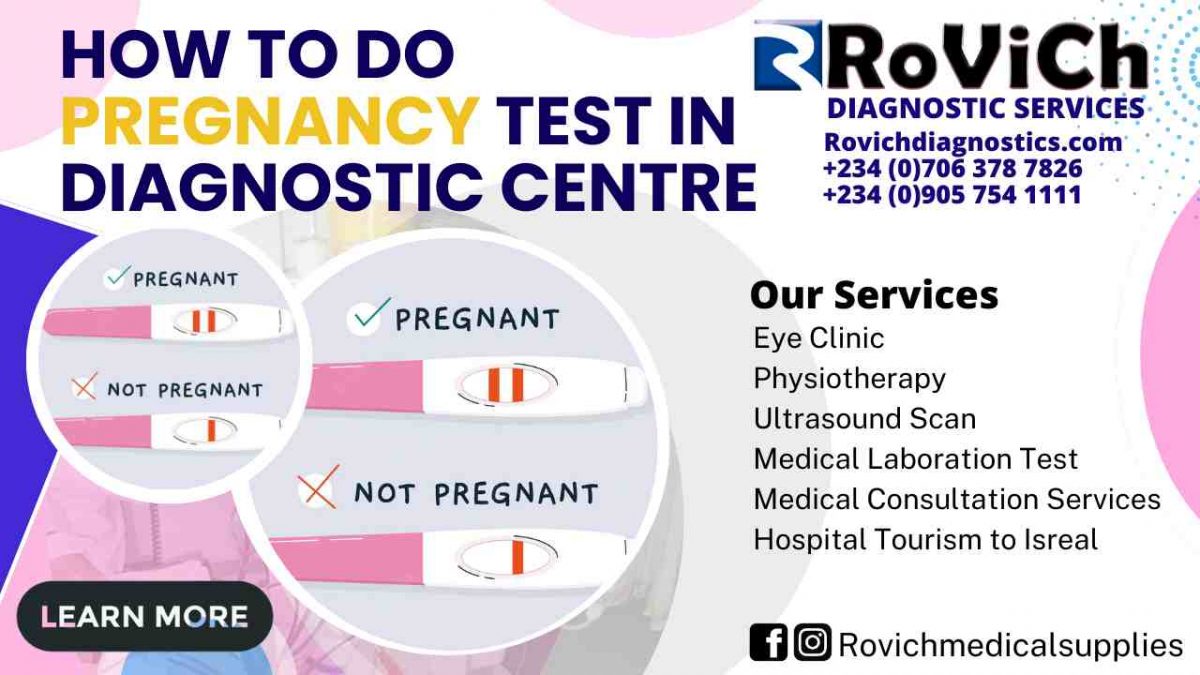
How-to-do-Pregnancy-Test-1200x675.jpg
30/Aug/2022

How to do Pregnancy Test

Introduction

Whether or whether you are pregnant may be determined with the use of a pregnancy test by determining the presence of a certain hormone in either your urine or blood. Human chorionic gonadotropin is the name of the hormone in question (HCG). After a fertilized egg has been implanted in a woman’s uterus, the placenta begins the process of producing HCG in the pregnant woman. In most cases, it is not produced at any other time except during pregnancy.

After you have gone without your period for about a week, you can take a urine pregnancy test to check for the presence of the HCG hormone. The test can either be performed at the office of a health care professional or at home with a test kit purchased separately. Because of the similarity between these tests, many women opt to take a pregnancy test they can do at home before seeing a medical professional. At-home pregnancy tests have an accuracy rate of between 97 and 99 percent when they are used appropriately.

What does it mean to take a pregnancy test?

A blood test for pregnancy is often performed in the office of a physician or other medical professional. It is able to detect lower quantities of HCG and can confirm or rule out a pregnancy earlier than a urine test. [Case in point:] [Case in point:] It is possible to determine whether or not you are pregnant with a blood test even before you have missed your period. Blood tests to determine pregnancy have an accuracy rate of around 99 percent. It is common practice to utilize a blood test to validate the findings of a pregnancy test that was performed at home.

What are some of its potential applications?

You can determine whether or not you are pregnant by taking a test specifically designed for that purpose.

Why do you need to take a pregnancy test?

If you suspect that you could be pregnant, you should get this test done. The signs and symptoms of pregnancy might differ from woman to woman, but the absence of a woman’s menstruation is typically the first indication that she is pregnant. A pregnancy may also be indicated by the following other frequent signs:

  • Breasts that are painful and swollen
  • Fatigue
  • Frequent urination
  • Feeling ill to one’s stomach and throwing up (also called morning sickness)
  • An uncomfortable feeling of fullness in the abdominal region

What exactly does it entail to take a pregnancy test?

There is no need for a doctor’s prescription in order to purchase a pregnancy test kit for at-home use. The vast majority are budget-friendly and simple to put into action.

A piece of equipment known as a dipstick is included in many different types of at-home pregnancy tests. Some of them also come with a cup for the collecting of donations. It’s possible that your at-home exam will involve the following stages, or actions very similar to them:

Urinate into the cup provided for the test immediately after you wake up in the morning. Because pee in the morning often has a higher concentration of HCG, the test results may be more reliable at this time.

Keep the dipstick in the stream of your urine for anywhere between five and ten seconds. Urinate into the collection cup that comes with the kit, and then place the dipstick into the cup for five to ten seconds. Kits that come with a collecting cup should be used in this manner.

Once you’ve waited a few minutes, the findings will be shown on the dipstick. The amount of time it takes to get results as well as the format in which the findings are displayed might differ significantly across different types of test kits.

Your dipstick may feature a window or another location that displays a plus or minus sign, a single or double line, or the words “pregnant” or “not pregnant.” Alternatively, it may simply read “not pregnant.” Instructions on how to interpret the findings of your pregnancy test will be included with the kit that you purchase.

If the test shows that you are not pregnant, you might wish to take it again in a few days. It’s possible that you took the test too soon in the pregnancy to get accurate results. During pregnancy, HCG levels steadily rise to higher and higher levels.


In the event that your test results indicate that you are pregnant, you need to schedule an appointment with your primary care physician as soon as possible. Your healthcare professional may choose to do a physical exam and/or a blood test in order to verify the findings of the previous tests.

A blood sample will be drawn from a vein in your arm by a qualified medical practitioner using a very thin needle. This will take place during a blood test. After the needle has been placed, a sample of the patient’s blood will be drawn into a test tube or a vial for further analysis. When the needle is inserted or removed, you could experience a brief yet sharp stinging sensation. In most cases, completion of this procedure will take less than five minutes.

Will there be anything specific that has to be done on my part to get ready for the test?

In order to do a pregnancy test on either your pee or your blood, you do not need to make any extra preparations.

Is there a possibility that the test might be harmful in some way?

There is no danger associated with getting a pee test done that anyone is aware of.

The possibility of something going wrong during a blood test is quite remote. You may experience some discomfort or bruising at the site where the needle was inserted, but for the most part, your symptoms will go away very fast.

What interpretations may be drawn from these results?

Depending on the results, you will learn whether or not you are pregnant. It is critical that you schedule an appointment with your primary care physician as soon as you discover that you are pregnant. There is a possibility that you will be referred to an obstetrician and gynecologist (OB/GYN) or a midwife, or that you may already be getting treatment from one of these medical professionals. These are medical professionals who concentrate on the wellbeing of women, prenatal care, and pregnancy as their primary areas of practice. During pregnancy, maintaining a healthy routine of doctor’s appointments and other preventative care is important for the health of both you and your unborn child.

you might be trying to get pregnant, and I was wondering if there’s anything else you should know about pregnancy tests.

A pregnancy test using urine can determine whether or not HCG is present in the body. The presence of HCG suggests that a pregnancy is present. The amount of HCG that is present can also be determined using a blood test for pregnancy. If your blood tests reveal a very low quantity of HCG, it is possible that you are experiencing an ectopic pregnancy. Ectopic pregnancies are those in which the pregnancy develops outside of the uterus. An ectopic pregnancy is not safe for the developing fetus since it occurs outside the uterus. In the absence of therapy, the illness can pose a significant risk of death to a woman.

Conclusion

We have been able to come to the knowledge in this article that you can take a urine pregnancy test to check for the presence of the HCG hormone. And that the test can either be performed at the office of a health care professional or at home with a test kit purchased separately. Because of the similarity between these tests, many women opt to take a pregnancy test they can do at home before seeing a medical professional. Fine the best diagnostic Centre where these are being done. Rovich Diagnostic Services

If you have any questions or concerns about what is written here please comment in the discussion below. contact us 

Hope this was helpful on the topic “How to do Pregnancy Test in Diagnostic Centre

Might also Interest you :  what are the symptoms of kidney diseases 

Might also Interest you: TYPES OF MEDICAL SCANNERS USED FOR DIAGNOSTIC IMAGING

Might also Interest you: DIFFERENCES BETWEEN HOSPITAL AND DIAGNOSTIC CENTRE 

Might also Interest you : How to leave healthy in a toxic environment

Might also Interest you : Ways to Reduce your high blood pressure   How to do Pregnancy Test

Might also Interest you : WHAT ARE THE CAUSES OF FIBROID How to do Pregnancy Test

Might also Interest you : Danger of self-medication  How to do Pregnancy Test

Might also Interest you : BEST MEDICAL EQUIPMENT FOR DIALYSIS IN 2022 How to do Pregnancy Test

Might also Interest you : How to stay free from color blindness How to do Pregnancy Test


Best-Ultrasound-scan-Centre-in-Awka-1200x675.jpg
29/Aug/2022

Best Ultrasound scan Centre in Awka

Introduction

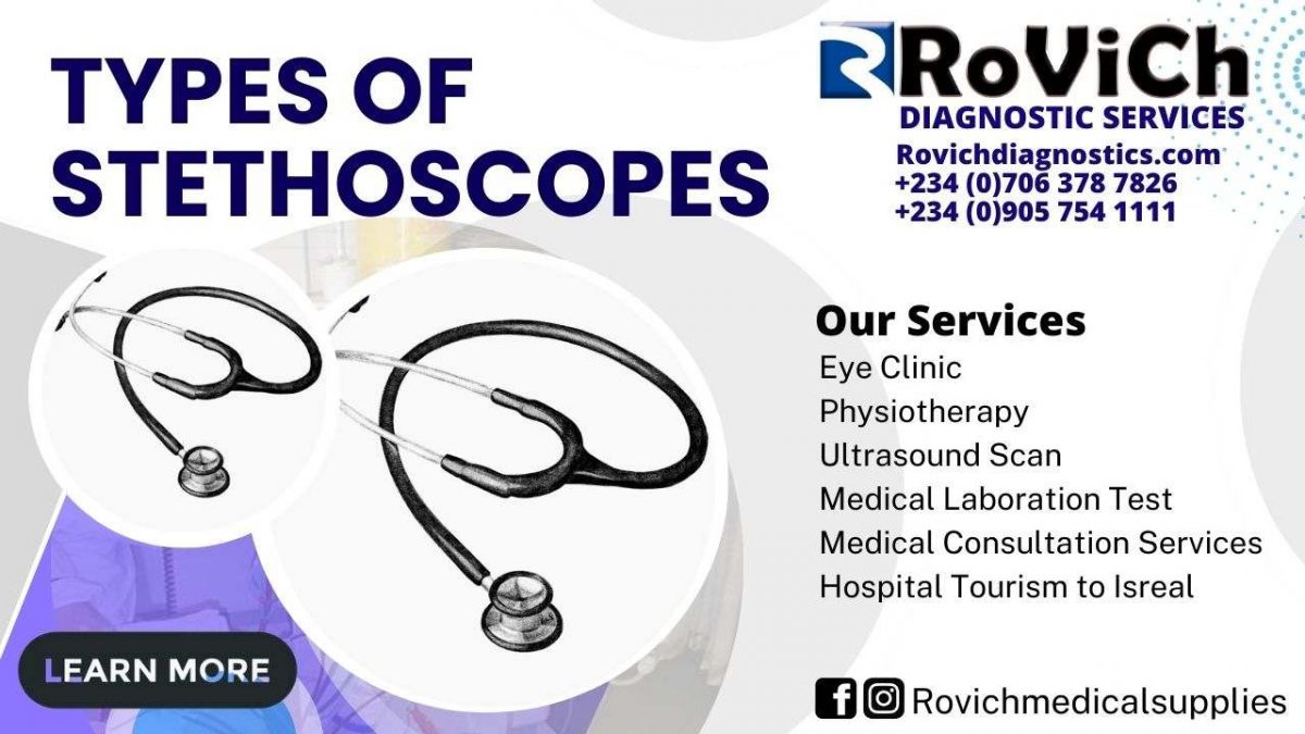
The auscultation, or listening to the inside noises of an animal or human body, is performed with a stethoscope, which is a piece of medical equipment. It normally consists of one or two tubes connected to two earpieces, as well as a small disc-shaped resonator that is worn against the skin and transmits sound. One can use a stethoscope to listen to the noises that are produced by the heart, lungs, or intestines, in addition to the sounds that are produced by the blood flow in the arteries and veins. When monitoring blood pressure, it is typically used in conjunction with a manual sphygmomanometer.

Less frequently, “mechanic’s stethoscopes,” which are equipped with rod-shaped chestpieces, are used to listen to the internal sounds made by machines (for instance, sounds and vibrations emitted by worn ball bearings), such as diagnosing a faulty automobile engine by listening to the sounds of its internal parts. Stethoscopes are commonly used to listen to the sounds of the heart and lungs. Stethoscopes have a variety of other uses, including checking scientific vacuum chambers for leaks and performing a variety of other small-scale acoustic monitoring activities. Stethoscopes were originally developed for medical usage.

Different kinds of stethoscopes

Fetal stethoscope

A fetal stethoscope, also known as a fetoscope, is a type of acoustic stethoscope that is formed in the shape of a listening horn. In order to listen to the heart beats of the fetus, it is put against the abdomen of a pregnant woman and held there. The fetal stethoscope, which was named after the French physician Adolphe Pinard, is also often called a Pinard horn (1844–1934).

3D-printed stethoscope

A 3D-printed stethoscope is an open-source medical gadget that is intended for auscultation and is created using 3D printing. Stethoscopes are used to listen to auscultation sounds.

Dr. Tarek Loubani, along with a group of medical and technological experts, was responsible for the creation of the 3D stethoscope. The design of the 3D-stethoscope, which was developed as part of the Glia project, is open source from the very beginning of the process. During the summer of 2015, the stethoscope received extensive coverage in the media.

Because of the blockade of the Gaza Strip, there was a shortage of stethoscopes and other essential medical equipment during the conflict in Gaza in 2012, which is when Loubani, a Palestinian-Canadian, was working as an emergency physician there. This prompted the need for a new type of stethoscope, which led to the development of the 3D-stethoscope. The Littmann Cardiology 3 stethoscope, which dates back to the 1960s, served as the inspiration for the 3D-printed stethoscope that was produced by Loubani.

Acoustic stethoscope

Acoustic stethoscopes accomplish their function by transmitting sound from the chest piece to the listener’s ears through hollow tubes that are filled with air. The chestpiece typically has two sides that can be placed against the patient for the purpose of sensing sound. These sides are known as the diaphragm (a plastic disc) and the bell (hollow cup). When the diaphragm is put on the patient, the patient’s body noises cause the diaphragm to vibrate, which results in the production of acoustic pressure waves. These waves then travel up the tube and are received by the listener. The vibrations of the patient’s skin directly produce acoustic pressure waves that travel up to the listener’s ears if the bell is put on the patient. The diaphragm is responsible for the transmission of higher frequency sounds, whereas the bell is responsible for the transmission of lower frequency sounds. The tube that connects into the chamber between the bell and the diaphragm can rotate and is only open on one side. This configuration ensures that the acoustic energy is sent primarily to either the bell or the diaphragm. When the opening is hooked into the bell, it may still be seen. The diaphragm can be connected to the tube by rotating it via the head by 180 degrees. In the early part of the 20th century, Rappaport and Sprague came up with the idea for the two-sided stethoscope that we use today.


Electronic stethoscope

The low sound levels can be solved by using an electronic stethoscope, also known as a stethophone, which electronically amplifies body noises. However, the amplification of stethoscope contact artifacts and component cutoffs (frequency response thresholds of electronic stethoscope microphones, pre-amps, amps, and speakers) limit the overall utility of electronically amplified stethoscopes. This occurs because electronically amplified stethoscopes amplify sounds in the middle frequency range while simultaneously dampening sounds in the high and low frequency ranges. Electronic stethoscopes are currently available from a variety of manufacturers. In order to achieve the best possible listening experience, electronic stethoscopes require the conversion of acoustic sound waves into electrical signals. These electrical impulses can then be amplified and analyzed. Electronic stethoscopes come with a wide variety of transducers, in contrast to acoustic stethoscopes, which all adhere to the same fundamental principles of physics. Putting a microphone in the chestpiece is the quickest way to accomplish sound detection, but it is also the method that is least accurate. Because of the interference that it causes from surrounding noise, this approach is no longer widely used. Another way is to position a piezoelectric crystal at the head of a metal shaft, with the bottom of the shaft making contact with a diaphragm. This technology is utilized in the Welch-Allyn Meditron stethoscope. In addition to this, 3M uses a piezoelectric crystal that is embedded in foam and hidden beneath a thick diaphragm that resembles rubber. In order to create a capacitive sensor, the Thinklabs Rhythm 32 makes use of an electromagnetic diaphragm that has a conductive inner surface. This particular diaphragm reacts to sound waves by causing changes in an electric field rather than reacting to changes in air pressure. Heart sounds can be wirelessly transmitted to a smartphone or tablet thanks to Eko Core’s capabilities.

An electronic stethoscope can be a wireless device, a recording device, and it can provide noise reduction, signal enhancement, and both visual and audio output because the sounds are sent electronically. Additionally, it can be a device that provides both visual and audio output. Around the year 2001, Stethographics introduced PC-based software that permitted the generation of a phonocardiograph, which is a graphic representation of cardiologic and pulmonologic sounds, and the interpretation of those sounds in accordance with related algorithms. For the goals of telemedicine (also known as remote diagnosis), as well as education, each of these aspects is helpful.

Electronic stethoscopes can also be used in conjunction with computer-aided auscultation applications to analyze recorded heart sounds and determine if the murmurs heard were diseased or harmless.

Doppler stethoscope

Electronic stethoscopes that assess the Doppler impact of ultrasound waves reflected from organs within the body are referred to as Doppler stethoscopes. The Doppler effect, which causes reflected waves to shift frequency, is used to determine the location of moving objects. Because of this, the Doppler stethoscope is ideally suited for use when dealing with moving targets, such as a heart that is beating. Recent research has shown that a continuous Doppler permits the auscultation of valve motions and blood flow noises in adults that are not audible when a heart examination is performed using a stethoscope. The traditional stethoscope auscultation had a sensitivity of 58%, but the Doppler auscultation had a sensitivity of 84%, when it came to the detection of aortic regurgitations. In addition, Doppler auscultation was superior in detecting reduced ventricular relaxation compared to conventional auscultation. Because the physics of traditional auscultation and Doppler auscultation are distinct from one another, it has been hypothesized that the two approaches could mutually benefit one another. A recently created Doppler-based military stethoscope that is immune to noise has been designed for the purpose of auscultation of patients in noisy surroundings.

Recording stethoscope

Some electronic stethoscopes have a direct audio output that may be connected to an external recording device like a laptop or an MP3 recorder. This allows the user to record their findings directly into the device. The same connection can be used to listen to the previously recorded auscultation through the stethoscope headphones, allowing for a more in-depth study for general research as well as evaluation and consultation regarding the condition of a specific patient, as well as telemedicine, also known as remote diagnosis.

There are applications available for smartphones that allow users to make the device function as a stethoscope. At least one of them makes advantage of the phone’s built-in microphone to amplify sound, create a visualization of it, and send the findings via e-mail. These applications may be utilized for the sake of teaching or as novelties; nevertheless, they have not yet acquired acceptability for use in professional medical settings. In 2015, the market saw the introduction of the first stethoscope that was compatible with a smartphone application.

Conclusion

We have been able to come to the knowledge in this article that a stethoscope is a piece of medical equipment used for auscultation, which is the process of listening to the sounds produced within the body of a person or animal. Fine the best diagnostic Centre where these are being used. Rovich Diagnostic Services

If you have any questions or concerns about what is written here please comment in the discussion below. contact us 

Hope this was helpful on the topic “Types of stethoscopes” Where to Buy Electrocardiogram Machines

Might also Interest you :  what are the symptoms of kidney diseases 

Might also Interest you: TYPES OF MEDICAL SCANNERS USED FOR DIAGNOSTIC IMAGING

Might also Interest you: DIFFERENCES BETWEEN HOSPITAL AND DIAGNOSTIC CENTRE 

Might also Interest you : How to leave healthy in a toxic environment Best Ultrasound scan Centre in Awka

Might also Interest you : Ways to Reduce your high blood pressure   Best Ultrasound scan Centre in Awka

Might also Interest you : WHAT ARE THE CAUSES OF FIBROID Best Ultrasound scan Centre in Awka

Might also Interest you : Danger of self-medication  Best Ultrasound scan Centre in Awka

Might also Interest you : BEST MEDICAL EQUIPMENT FOR DIALYSIS IN 2022 Best Ultrasound scan Centre in Awka

Might also Interest you : How to stay free from color blindness Best Ultrasound scan Centre in Awka


Where-to-Buy-Electrocardiogram-Machines-1200x675.jpg
26/Aug/2022

Where to Buy Electrocardiogram Machines

Introduction

Did you know that cardiovascular disease continues to be the leading cause of mortality in the United States? Each year, heart attacks affect over 800,000 people in the United States.

Because of this, there is a rise in the number of people who become disabled. Electrocardiogram machines are helpful in identifying cardiac abnormalities that can point to an increased risk of developing heart disease. In addition to that, it is the method of choice for diagnosing heart attacks.


Continue reading if you are curious about the inner workings of these machines and want to find out more about how they function.


What Is an Electrocardiogram and How Does It Work?

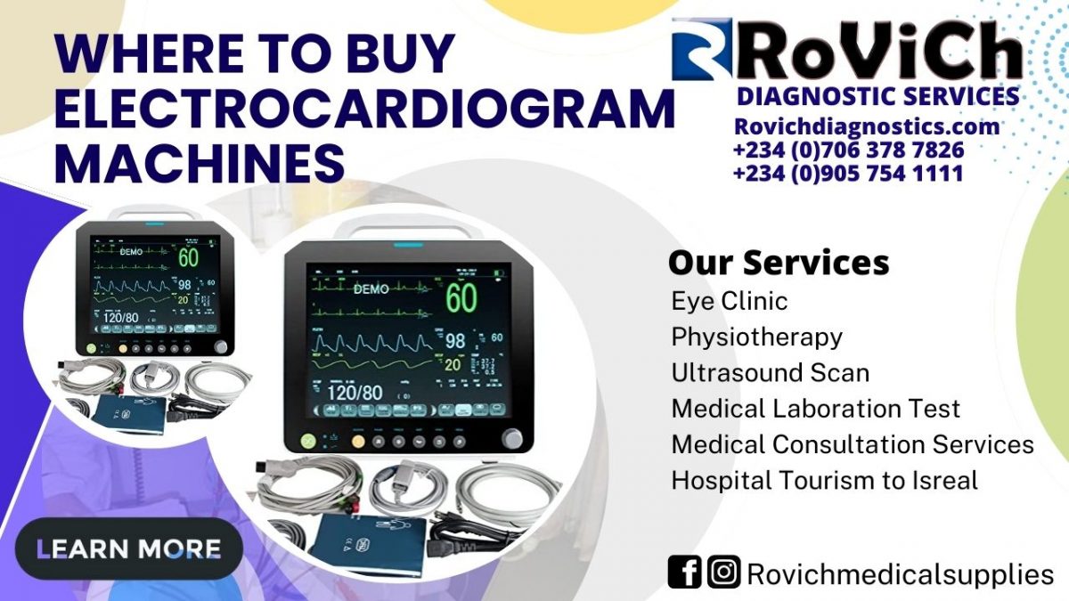
An ECG is primarily used to record and analyze the electrical activity coming from the heart. This is accomplished by having a variety of electrodes attached to the torso region by a trained professional. Electrodes are able to take up signals whenever your heart contracts and blood is pumped.


ECGs can reveal a variety of information, including the following:

  • Abnormal rhythms of the heart
  • Heart attack
  • the capacity of your heart
  • spaces within the heart
  • Heart injury

Inpatient hospital settings can benefit from the usage of ECGs. Patients who suddenly complain of chest discomfort, arrhythmias, or dizziness are candidates for treatment with these devices. These tests are given to patients who are currently being treated for heart problems by their physicians.


Patients who are at an increased risk of having a heart disease can benefit from their use. In addition, they are typically used for monitoring a patient’s pacemaker, which is a device that helps manage the electrical component of your heartbeat and your heart rhythm.


A brief electrocardiogram might provide your surgeon with valuable information regarding your heart health prior to an anticipated surgical procedure. In exchange, this can help determine whether or not you are at a greater risk for getting general anesthesia.


Electrocardiogram Test

How precisely does one use an electrocardiogram? To begin, a Holter monitor may be recommended by a medical professional in the event that you do not exhibit ongoing symptoms. Over the course of one to two days, this will be able to monitor the activity of your heart.


You can carry it with you to your house because it is portable. A regular electrocardiogram machine will monitor the heart event as it is taking place. Your torso and either your upper or lower limbs each have one of twelve distinct leads linked to them. At this point, the patient needs to take off any jewelry and clothing that is covering their upper body.


Following this step, the leads are reconnected to the monitor. During this test, you will most likely be required to lay down, and in some areas of your head, you may be asked to shave sections of hair in order to establish a proper connection.


During the time that the exam is being administered to you, you are exempt from performing any tasks. Your heart will be monitored by the electrocardiogram machine for a predetermined amount of time, after which the examination will be finished.


Changing the position of the electrodes or touching them while the test is ongoing has the potential to skew the results. In order to get the most precise readings possible, it is essential that the individual lie absolutely still on the table.


Your cardiologist will be able to determine whether or not there is anything wrong with your heart based on the findings of the ECG test. If the results are abnormal or cannot be determined, then it is possible that additional test will be necessary. In addition to this, additional follow-up testing might be performed using an echocardiography.


Interpreting ECG Results

The results are interpreted by medical professionals by comparing the ECG printout to the range of normal readings. The reading presents information regarding the beat of the heart.


There are three primary components to it:

  • P wave
  • QRS
  • T wave

The amplitude is a measurement that indicates how high each wave is. For instance, the height of the P wave, on average, is between between 2 and 2.5 millimeters tall. The number of squares that it reaches on the table is the metric that practitioners use to measure it.


In this particular illustration, it is two and a half squares. The length of time that an ECG is recorded for is yet another common feature that medical professionals investigate. You may take another look at the P wave as an example.


The P wave lasts anywhere from 0.06-0.12 seconds on average. This range of time is considered to be its normal length. When these results in the P wave are aberrant, it may suggest that the atriums have grown in size. Each segment of the electrocardiogram provides clues as to what the potential issue could be.


ECG Versus Vital Sign Monitors

What sets an electrocardiogram apart from other types of monitors used to check vital signs? Electrocardiogram machines are used for the exclusive purpose of analyzing heart rhythms. The sensitivity and accuracy of this test are superior to those of blood pressure and pulse examinations.


A standard monitor for vital signs is able to evaluate a number of various vitals, including the following:

  • Oxygen saturation (SpO2)
  • Pulse rate
  • The rate of respiration
  • Blood pressure
  • End-tidal carbon dioxide (ETCO2)

Although some of these machines are capable of performing straightforward ECG readings, their primary function is to monitor patients and do various vital assessments. It is not intended to treat specific cardiac diseases on a regular basis and hence cannot do so.

Inpatient medical institutions typically feature a monitor for vital signs and are able to bring in an electrocardiogram machine if one is required.

Technology that can be worn

The use of portable technology for the purpose of monitoring one’s health has gained widespread popularity. In one piece of research, the accuracy of ECG readings taken on a watch or other portable device was compared to that of traditional ECG machines.


The Apple Watch was the primary topic of investigation for this study. There have been advertising for products that can assist with the diagnosis of atrial fibrillation using photoplethysmography (PPG).


This technology is able to monitor changes in blood volume through the use of light sensors that are embedded inside the watch band. It is a single lead ECG readout that has Class II clearance from the Food and Drug Administration.


This indicates that it should not be used as a conclusive diagnosis at any point in time. Instead, extreme caution needs to be exercised whenever these measurements are used to diagnose atrial fibrillation. It is strongly recommended that you get in touch with a qualified medical professional.


The Apple Watch had a sensitivity of roughly 96% when it was used to detect atrial fibrillation, and it had a specificity of approximately 97% when it was used in this capacity. The risk of producing false positives or negatives is the primary worry raised by the use of wearable technologies.


In addition to this, there is a rising risk that individuals will quit wearing it after a few months to a year has passed. This presents a one-of-a-kind obstacle in the process of treating atrial fibrillation. This heart issue is a chronic illness that calls for ongoing monitoring and treatment.


It is also not as accurate as readings taken from an ECG with 12 leads because this one only has one lead.


12-Lead ECG Studies

Other research have investigated the ways in which medical professionals can achieve a higher level of precision while interpreting 12-lead ECG results. Despite breakthroughs in technology, there is still room for improvement in artificial intelligence in this domain.


ECGs provide a number of challenges, one of which is the need for a cardiologist to interpret the results. Readings from a single lead electrocardiogram were the first step in the development of artificial intelligence; a prime example of this may be found when you use your Apple Watch to perform a short screening.


At this point in time, these algorithms have not been adequately translated to 12-lead readings. In this study, the use of technology to interpret the results of a 12-lead electrocardiogram was investigated.


The electrocardiogram (ECG) can frequently reveal more than one heart problem. The majority of cardiologist reports, on the other hand, exclusively categorize a single condition, such as atrial fibrillation.


When it came to the identification of heart problems, the researchers discovered that automated systems gave results that were relatively accurate. The researchers had high hopes that one day there will be access to a larger, open-source database that contains electrocardiogram (ECG) images.


According to the findings of this study, accurate ECG readings are the key to advancing cardiology and the diagnosis of medical disorders.


There are already electrocardiograms on the market that provide software for ECG interpretation. Because they assist cardiologists in making diagnosis, these machines are among the most effective ones now available on the market.


It provides a more comprehensive database for analyzing normal versus aberrant rhythms and comparing the two. It also has the capability to retain this information as well as the data from the ECG, allowing you to transfer it to other facilities or units.


ECGs Have More Than One Use

Electrocardiograms are not just utilized in cardiology clinics but also in other medical settings. They are useful in a variety of settings, ranging from intensive care units to outpatient clinics. Additionally, they are utilized in end-of-life care provision.


In intensive care units, the use of ECGs is mandatory; yet, they frequently result in slower response times on the part of nurses and practitioners. This is because the majority of ECGs come equipped with some sort of warning system.


This condition has been given the name “alarm tiredness” by researchers and medical professionals. How do you go about counteracting this?


Electrocardiogram equipment are intended for monitoring of the highest possible sensitivity. Because of this, it is likely that not all of the alarms will be required in every sort of environment. Only 11 of the 151 alarms that occurred during end-of-life care were judged to be indicative of atrial fibrillation, according to the findings of the research.


On the other hand, over three quarters of the alarms were audible, which necessitated the presence of a nurse or other member of the medical staff in the room. In this particular context, providing the patient and their family with comfort care is the primary focus.


It is possible that intensive care units require audible alarms whenever there is even a slight change in the electrocardiogram (ECG), although it is highly unlikely that this setting requires such alarms. This demonstrates the importance of having ECG machines that have a variety of adjustable settings.


It is essential that you invest in ECGs that are able to accommodate a variety of patients, environments, and diagnoses. For instance, the frequency and detection ranges are going to be different for a juvenile patient than they are going to be for an adult patient.


Rather than investing in a number of different kinds of electrocardiograms (ECGs), your medical facility would benefit financially and operationally from owning just one that can perform all of the necessary functions.


Electrocardiogram Equipment of the Highest Quality

You are already familiar with the distinctions, benefits, and drawbacks that exist between a single-lead and a 12-lead electrocardiogram (ECG). There are a lot of different kinds of ECG equipment available on the market nowadays.


An electrocardiogram (ECG) machine with two leads and one with six leads are other common types. A reading taken from an electrocardiogram with six leads is considered to be the next best thing to one with 12 leads.


Recent research has shown that it is more accurate than your Apple Watch, but it is still not quite as exact as the method that has been used for decades. Instead, it is more likely to be utilized for self-monitoring and bringing awareness to cardiovascular issues.


A 12-lead ECG machine is still the most accurate option for diagnosing cardiac conditions definitively.

Where to Buy Electrocardiogram Machines

Electrocardiogram equipment are crucial components of any hospital or medical center. They are able to provide practitioners with a concise insight into the cardiac and general health of a patient. Click here to Buy today  Rovich Medical Supplies or Choose any of the listed Electrocardiogram equipment Below:

Conclusion

We have been able to come to the knowledge in this article that Electrocardiograms are not just utilized in cardiology clinics but also in other medical settings. They are useful in a variety of settings, ranging from intensive care units to outpatient clinics.… Fine the best diagnostic Centre for all type of Test at Rovich Diagnostic Services

If you have any questions or concerns about what is written here please comment in the discussion below. contact us 

Hope this was helpful on the topic “What is the Function of an Electrocardiogram Machine?” Where to Buy Electrocardiogram Machines

Might also Interest you :  what are the symptoms of kidney diseases 

Might also Interest you: TYPES OF MEDICAL SCANNERS USED FOR DIAGNOSTIC IMAGING

Might also Interest you: DIFFERENCES BETWEEN HOSPITAL AND DIAGNOSTIC CENTRE 

Might also Interest you : BEST MEDICAL EQUIPMENT FOR DIALYSIS IN 2022 Where to Buy Electrocardiogram Machines

Might also Interest you : How to stay free from color blindness Where to Buy Electrocardiogram Machines

Might also Interest you : How to leave healthy in a toxic environment Where to Buy Electrocardiogram Machines

Might also Interest you : Ways to Reduce your high blood pressure   Where to Buy Electrocardiogram Machines

Might also Interest you : WHAT ARE THE CAUSES OF FIBROID Where to Buy Electrocardiogram Machines

Might also Interest you : Danger of self-medication  Where to Buy Electrocardiogram Machines


How-to-reduce-blood-pressure-Rovich-Diaganostic-Services-1200x675.jpg
25/Aug/2022

How to reduce blood pressure

Watch your weight and waistline.

Weight increases blood pressure. Overweight causes sleep apnea, which elevates blood pressure.

Losing weight is an effective way to lower blood pressure. Even a minor weight loss can reduce blood pressure in overweight or obese people. Each kilogram (2.2 pounds) decreased may lower blood pressure by 1 mm Hg.

Also, waistline matters. Too much belly fat might raise blood pressure.

Typically:

Men with a 40-inch waist are at risk (102 centimeters).
Women with a 35-inch waist are at risk (89 centimeters).
Ethnic groups’ numbers vary. Request a waist measurement from your doctor.

Regularly exercise

Regular exercise can reduce hypertension by 5 to 8 mm Hg. Keep exercising to prevent high blood pressure. Aim for 30 minutes of moderate daily exercise.

Exercise helps prevent high blood pressure from developing (hypertension). Regular exercise can lower hypertensives’ blood pressure.

Walking, jogging, cycling, swimming, and dancing can reduce blood pressure. Then there’s HIIT. This workout alternates hard bursts with gentler times.

Weightlifting lowers blood pressure. At least twice a week, including strength training. Consult a doctor about a workout plan.

Eat well

A diet rich in whole grains, fruits, vegetables, and low-fat dairy products can lower blood pressure by 11 mm Hg. The DASH and Mediterranean diets help reduce blood pressure.

Potassium reduces the blood pressure effects of sodium. Fruits and vegetables are better than supplements for potassium. 3,500 to 5,000 mg a day may reduce blood pressure 4 to 5 mm Hg. Ask your doctor about potassium.

Eat less salt

Even a little reduction in salt can enhance heart health and lower blood pressure by 5-6 mm Hg.

Sodium intake affects blood pressure differently among groups. Limit daily salt to 2,300 mg. Most adults should consume 1,500 mg or less of sodium daily.

To cut sodium:

Labels matter. Choose low-sodium foods and drinks.
Reduce processed foods. Foods naturally have little sodium. Processing adds most sodium.
Salt-free. Flavor food using herbs and spices.
Cook. Cooking reduces sodium levels.

Reduce drinking

Less than one drink a day for women or two for males can reduce blood pressure by 4 mm Hg. 12 ounces of beer, 5 ounces of wine, or 1.5 ounces of 80-proof liquor equals one drink.

Too much alcohol raises blood pressure. It reduces blood pressure drugs’ effectiveness.

Tobacco-free

Smoking raises BP. Cigarette cessation lowers blood pressure. It can minimize heart disease risk, improve overall health, and extend life.

Sleep well

Less than six hours of sleep per night for weeks might cause hypertension. Sleep apnea, RLS, and sleepiness can interrupt sleep (insomnia).

Describe your sleep problems to your doctor. Treating the reason can assist sleep. If you don’t have sleep apnea or RLS, try these sleep suggestions.

Schedule your sleep. Keep a consistent sleep-wake schedule. Keep a consistent weeknight and weekend schedule.
Relax. That indicates cool, calm, dark. Before bed, relax. Warm baths and relaxation techniques may help. Avoid TV and computer screen light.
Eat and drink carefully. Don’t sleep hungry or full. Eat light before bed. Limit nicotine, coffee, and alcohol before bed.
Nap less. Limiting daytime naps to 30 minutes may aid nocturnal sleep for nappers.

Relax

Chronic stress can raise blood pressure. Stress reduction approaches may lower blood pressure, but further research is needed.

Determine what generates stress, such as work, family, finances, or sickness, and eliminate it. Try:

Don’t overcommit. Prioritize your day. Yes, but. Allow enough time for tasks.
Plan to solve challenges you can control. Supervisors can help with job issues. Find strategies to resolve family conflicts.
Reduce stress. If rush-hour traffic stresses you out, consider taking public transit. Avoid stress-causing folks.
Unwind. Sit quietly and breathe deeply daily. Take a walk, cook, or volunteer.
Thankfulness. Gratitude reduces stress.

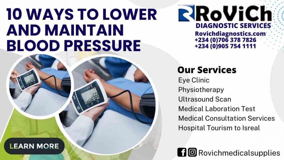
Regularly check your blood pressure at home.

Blood pressure can be monitored at home. It can test your drugs and lifestyle modifications.

Home blood pressure monitors are nonprescription. Consult a doctor before starting home monitoring.

Controlling blood pressure requires regular doctor visits. Ask your provider how often to check your blood pressure if it’s well-controlled. You may only need to check it daily.

Aid

Healthy people have supportive family and friends. They may encourage you to take care of yourself, drive you to the doctor, or start an exercise regimen with you.

Join a support group if you need help beyond family and friends. This may put you in touch with people who can boost your mood and offer coping tips.


Conclusion

We have been able to come to the knowledge in this article that a diet rich in whole grains, fruits, vegetables, and low-fat dairy products can lower blood pressure by 11 mm Hg. and that the DASH and Mediterranean diets help reduce blood pressure… Fine the best diagnostic Centre for all type of Blood Test done at Rovich Diagnostic Services

If you have any questions or concerns about what is written here please comment in the discussion below. contact us 

Hope this was helpful on the topic “10 ways to lower and maintain blood pressure.

Might also Interest you :  what are the symptoms of kidney diseases 

Might also Interest you: TYPES OF MEDICAL SCANNERS USED FOR DIAGNOSTIC IMAGING

Might also Interest you: DIFFERENCES BETWEEN HOSPITAL AND DIAGNOSTIC CENTRE

Might also Interest you : BEST MEDICAL EQUIPMENT FOR DIALYSIS IN 2022 Being a Universal Blood Donor Rovich

Might also Interest you : How to stay free from color blindness Being a Universal Blood Donor Rovich

Might also Interest you : How to leave healthy in a toxic environment Being a Universal Blood Donor Rovich

Might also Interest you : Ways to Reduce your high blood pressure   Being a Universal Blood Donor Rovich

Might also Interest you : WHAT ARE THE CAUSES OF FIBROID Being a Universal Blood Donor Rovich

Might also Interest you : Danger of self-medication  Being a Universal Blood Donor Rovich


How-to-safeguard-myself-against-HIV-1200x675.jpg
25/Aug/2022

HIV can be transmitted through a woman’s sperm, blood, vaginal fluids, and breast milk. Protect yourself by always using a condom when engaging in sexual activity, and never sharing needles with anyone else. You can also discuss PrEP, which is a daily drug that helps prevent HIV, with your primary care physician.

How can I protect myself from contracting HIV when having sex?

HIV is transmitted by sexual intercourse, specifically vaginal and anal sex, as well as through contact with blood or sexual fluids (such as sperm and vaginal fluids). Therefore, the only method to absolutely guarantee that one will not contract HIV is to abstain from engaging in sexual activity of any kind.

However, the majority of people do engage in sexual activity at some time in their lives, which is why it is essential to have knowledge regarding HIV prevention and know how to engage in sexual activity in a safer manner. The risk of contracting HIV can be significantly reduced by using condoms. The most effective method of preventing the spread of HIV is to use a condom every single time you engage in sexual activity with another person. In addition, there is a daily tablet that you can take that is referred to as PrEP, and it can help prevent HIV. If you ask your doctor or nurse, they should be able to tell you whether or not PrEP is right for you.


When it comes to the risk of contracting HIV, certain sexual practices are less risky than others. These behaviors are considered “low risk” because there have never been any documented cases of HIV transmission linked to them:

  • masturbating
  • genital contact between partners is not appropriate.
  • putting your bodies up against one another (dry humping)
  • kissing
  • oral sexual activity while wearing a condom or dental dam
  • using only sanitary objects for sexual activity

These behaviors are considered to have a “reduced risk” since they have only been linked to a small number of documented cases of HIV (out of millions):

  • “French” or deep kissing (if the person with HIV has sores or bleeding in their mouth)
  • sexual contact using a condom and/or other preventative measures
  • anal sex while using a condom and/or other forms of PrEP
  • oral intercourse that is not protected by a dental dam or condom

These behaviors are considered “high risk” because they are responsible for the transmission of HIV to millions of people:

  • sexual contact in the genital area without using a condom or another kind of PrEP
  • sexual activity of a non-condomed, non-PrEP nature.


It is much simpler for HIV to enter your body if you have cuts, sores, or other areas in your skin through which blood, vaginal fluids, or sperm could potentially enter. This is because HIV is transmitted through sexual contact. If you are experiencing an outbreak of herpes or any other virus, you should avoid having sexual contact. It is recommended that you get tested for sexually transmitted diseases (STDs) on a regular basis because having other STDs increases your risk of getting HIV.


There is now no vaccine available that can protect against HIV, although many people are working hard to develop one. Also, there are medicines that can help prevent HIV, and they go by the names PEP and PrEP.


Antiretroviral therapy is a form of HIV treatment that can be administered to your partner even if you yourself do not carry the virus (ART). They have a lower risk of transmitting HIV through sexual activity if they are receiving ART. In addition, some persons who are receiving ART are unable to pass the virus on to their partners at all.


What exactly is pre-exposure prophylaxis, and how does it prevent HIV infection?

The term “pre-exposure prophylaxis” is abbreviated as “PrEP.” It is a medication that you take once a day to help prevent HIV. You only need to take it once. Your doctor or nurse can help you determine if PrEP works sense for you. Learn more about the PrEP program.


What exactly is pre-exposure prophylaxis (PEP), and how does it protect against HIV?

Post-exposure prophylaxis is what we refer to as PEP. Your risk of contracting HIV is reduced by starting a course of medication called antiretroviral therapy (ART) after you have been exposed to HIV. In order for pre-exposure prophylaxis (PEP) to be effective, it must be initiated within 72 hours (or three days) of an individual’s initial HIV infection. The sooner you get started, the better off you will be. Because every hour counts, you should get checked out as soon as possible if you have any reason to believe that you may have been exposed to HIV. Call your nurse or doctor, or go to the emergency hospital. PEP is only meant to be used in life-threatening situations; it is not a substitute for wearing condoms or PrEP. Learn more about PEP by reading.


What exactly is antiretroviral therapy (ART), and how does it work to stave off HIV?

Antiretroviral therapy, also known as ART, refers to a regimen of medications that, when taken together, mitigate the harmful effects of HIV on the body. If you take these medications, you may be able to maintain your health for many years. Additionally, it can lessen or even eliminate the possibility that you will pass HIV on to another person.


Your HIV count, also known as your viral load, will drop significantly as a result of taking ART, often to the point where it will not be detectable with conventional blood testing. It is referred to as being “undetectable” when a person’s HIV viral load is so low that some tests are unable to detect it. When a person’s viral load is undetectable, they are unable to transmit HIV to other people through sexual contact with them.


It is essential to keep in mind that even if your viral load is undetectable, HIV is still active in your body. This fact cannot be stressed enough. If you stop taking HIV medication, your viral load may increase, which increases the risk that you will spread HIV on to others through sexual contact. Finding the treatment that works best for you to help keep your viral load as low as possible and maintain your health is something that your doctor or nurse can assist you in doing.


How can I protect myself from sexually transmitting HIV to another person?

If you find out that you have HIV, it is important that you maintain your composure. People with HIV can lead normal, healthy sexual lives and have normal, healthy relationships with others. However, it is critical to exercise caution in order to assist your partner(s) in avoiding contracting HIV.

There are a few things you can do to keep from passing the HIV virus on to other people, including the following:

  • Always wear condoms, whether you’re having an oral or a vaginal sexual encounter.
  • You should begin HIV therapy as soon as possible, and you should continue taking your HIV medication. When administered correctly, HIV therapy has the potential to significantly reduce or even eliminate the risk of transmitting the virus to a person’s subsequent sexual partners (and help you stay healthy).
  • PrEP is a daily tablet that can reduce the likelihood of your spouse contracting HIV. They can take it together with you.
  • Never share needles that you use for injecting narcotics, getting pierced, or tattooed.
  • Regularly get tested for and treated for other sexually transmitted diseases (STDs) in addition to HIV. It is more simpler to pass on HIV to other people if you already have one or more other STDs.

If you have tested positive for HIV, it is vital that you inform all of your sexual partners of this fact so that they can also get tested for the virus. Even if you take all of the necessary precautions to prevent the spread of HIV, you should still be open and honest with your future partners about your status. This will allow you both to be educated and better assist one another in maintaining a healthy lifestyle.

Conclusion

We have been able to come to the knowledge in this article that always wearing condoms, whether you’re having an oral or a vaginal sexual encounter will help to reduce the spread of HIV from a partner. it is also good to begin HIV therapy as soon as possible, and you should continue taking your HIV medication because when administered correctly, HIV therapy will significantly reduce or even eliminate the risk of transmitting the virus to a person’s subsequent sexual partners. Fine the best diagnostic Centre for HIV test at Rovich Diagnostic Services

If you have any questions or concerns about what is written here please comment in the discussion below. contact us 

Hope this was helpful on the topic “Exactly what steps can I take to safeguard myself against HIV?

Might also Interest you :  what are the symptoms of kidney diseases 

Might also Interest you: TYPES OF MEDICAL SCANNERS USED FOR DIAGNOSTIC IMAGING

Might also Interest you: DIFFERENCES BETWEEN HOSPITAL AND DIAGNOSTIC CENTRE

Might also Interest you : BEST MEDICAL EQUIPMENT FOR DIALYSIS IN 2022 How to safeguard myself against HIV

Might also Interest you : How to stay free from color blindness How to safeguard myself against HIV

Might also Interest you : How to leave healthy in a toxic environment How to safeguard myself against HIV

Might also Interest you : Ways to Reduce your high blood pressure   How to safeguard myself against HIV

Might also Interest you : WHAT ARE THE CAUSES OF FIBROID How to safeguard myself against HIV

Might also Interest you : Danger of self-medication  How to safeguard myself against HIV


Rovich medical Services is a top diagnostic centre that provides world class medical services


Social networks


Facebook

www.facebook.com


Twitter

#WeCare


Instagram

www.instagram.com


Emergency number


0905 754 1111


Call us now if you are in a emergency laboratory need, we will reply swiftly and provide you with an aid


Copyright by Rovich Medical Services. All rights reserved.