Blog

Completely synergize resource taxing relationships via premier niche markets. Professionally cultivate one-to-one customer service with robust ideas.
Blue-Controller-Faux-OrganicAbstract-Technology-Gaming-Facebook-Shops-Cover-25-1200x900.jpg
08/Sep/2021

Best place for diabetes Diagnosis

INTRODUCTION

Diabetes mellitus (DM), commonly known as just diabetes, is a group of metabolic disorders characterized by a high blood sugar level over a prolonged period of time. Symptoms often include frequent urination, increased thirst, and increased appetite. If left untreated, diabetes can cause many health complications. Acute complications can include diabetic ketoacidosis, hyperosmolar hyperglycemic state, or death. Serious long-term complications include cardiovascular disease, stroke, chronic kidney disease, foot ulcers, damage to the nerves, damage to the eyes, and cognitive impairment.

Diabetes is due to either the pancreas not producing enough insulin or the cells of the body not responding properly to the insulin produced. There are three main types of diabetes mellitus. Also is a condition in which blood glucose levels are too high to be considered normal but not high enough to be labeled diabetes. People have prediabetes if their fasting blood glucose level is between 100 mg/dL (5.6 mmol/L) and 125 mg/dL (6.9 mmol/L) or if their blood glucose level 2 hours after a glucose tolerance test is between 140 mg/dL (7.8 mmol/L) and 199 mg/dL (11.0 mmol/L). Prediabetes carries a higher risk of future diabetes as well as heart disease. Decreasing body weight by 5 to 10% through diet and exercise can significantly reduce the risk of developing future diabetes. The three major nutrients that make up most food are carbohydrates, proteins, and fat. Sugars are one of three types of carbohydrates, along with starch and fiber.

There are many types of sugar. Some sugars are simple, and others are complex. Table sugar (sucrose) is made of two simpler sugars called glucose and fructose. Milk sugar (lactose) is made of glucose and a simple sugar called galactose. The carbohydrates in starches, such as bread, pasta, rice, and similar foods, are long chains of different simple sugar molecules. Sucrose, lactose, carbohydrates, and other complex sugars must be broken down into simple sugars by enzymes in the digestive tract before the body can absorb them. Once the body absorbs simple sugars, it usually converts them all into glucose, which is an important source of fuel for the body. Glucose is the sugar that is transported through the bloodstream and taken up by cells. The body can also make glucose from fats and proteins. Blood “sugar” really means blood glucose.

Best place for diabetes diagnosis

TYPES OF DIABETES

Type 1 diabetes:

In type 1 diabetes (formerly called insulin-dependent diabetes or juvenile-onset diabetes), the body’s immune system attacks the insulin-producing cells of the pancreas, and more than 90% of them are permanently destroyed. The pancreas, therefore, produces little or no insulin. Only about 5 to 10% of all people with diabetes have type 1 disease. Most people who have type 1 diabetes develop the disease before age 30, although it can develop later in life.  Scientists believe that an environmental factor—possibly a viral infection or a nutritional factor during childhood or early adulthood—causes the immune system to destroy the insulin-producing cells of the pancreas. A genetic predisposition makes some people more susceptible to environmental factors.

Medical Consultation

Type 2 diabetes:  In type 2 diabetes (formerly called non– insulin-dependent diabetes or adult-onset diabetes), the pancreas often continues to produce insulin, sometimes even at higher-than-normal levels, especially early in the disease. However, the body develops resistance to the effects of insulin, so there is not enough insulin to meet the body’s needs. As type 2 diabetes progresses, the insulin-producing ability of the pancreas decreases. Type 2 diabetes was once rare in children and adolescents but has become more common. However, it usually begins in people older than 30 and becomes progressively more common with age. About 26% of people older than 65 have type 2 diabetes. People of certain racial and ethnic backgrounds are at increased risk of developing type 2 diabetes: blacks, Asian Americans, American Indians, and people of Spanish or Latin American ancestry who live in the United States have a twofold to threefold increased risk as compared with whites. Type 2 diabetes also tends to run in families. Obesity is the chief risk factor for developing type 2 diabetes, and 80 to 90% of people with this disorder are overweight or obese. Because obesity causes insulin resistance, obese people need very large amounts of insulin to maintain normal blood glucose levels. Certain disorders and drugs can affect the way the body uses insulin and can lead to type 2 diabetes.  Examples:  of common states (conditions) that result in impaired insulin use are High levels of corticosteroids (most commonly due to use of corticosteroid drugs or Cushing syndrome Pregnancy (gestational diabetes ) Diabetes also may occur in people with excess production of growth hormone (acromegaly ) and in people with certain hormone-secreting tumors. Severe or recurring pancreatitis and other disorders that directly damage the pancreas can lead to diabetes.

 Contact the Best place for diabetes Diagnosis and find out more Causes of diabetes

Diabetes mellitus is classified into six categories: type 1 diabetes, type 2 diabetes, hybrid forms of diabetes, hyperglycemia first detected during pregnancy, “unclassified diabetes”, and “other specific types”. The “hybrid forms of diabetes” contains slowly evolving, immune-mediated diabetes of adults and ketosis-prone type 2 diabetes. The “hyperglycemia first detected during pregnancy” contains gestational diabetes mellitus and diabetes mellitus in pregnancy (type 1 or type 2 diabetes first diagnosed during pregnancy). The “other specific types” are a collection of a few dozen individual causes. Diabetes is a more variable disease than once thought and people may have combinations of forms. The term “diabetes”, without qualification, refers to diabetes mellitus.

Rovich medical supplies

Type 1

Type 1 diabetes is characterized by loss of the insulin-producing beta cells of the pancreatic islets, leading to insulin deficiency. This type can be further classified as immune-mediated or idiopathic. The majority of type 1 diabetes is of an immune-mediated nature, in which a T cell-mediated autoimmune attack leads to the loss of beta cells and thus insulin. It causes approximately 10% of diabetes mellitus cases in North America and Europe. Most affected people are otherwise healthy and of a healthy weight when onset occurs. Sensitivity and responsiveness to insulin are usually normal, especially in the early stages. Although it has been called “juvenile diabetes” due to the frequent onset in children, the majority of individuals living with type 1 diabetes are now adults. Contact the Best place for diabetes diagnosis Rovich Diagnostics Services 

“Brittle” diabetes, also known as unstable diabetes or labile diabetes, is a term that was traditionally used to describe the dramatic and recurrent swings in glucose levels, often occurring for no apparent reason in insulin-dependent diabetes. This term, however, has no biologic basis and should not be used. Still, type 1 diabetes can be accompanied by irregular and unpredictable high blood sugar levels, and the potential for diabetic ketoacidosis or serious low blood sugar levels. Other complications include an impaired counterregulatory response to low blood sugar, infection, gastroparesis (which leads to erratic absorption of dietary carbohydrates), and endocrinopathies (e.g., Addison’s disease).These phenomena are believed to occur no more frequently than in 1% to 2% of persons with type 1 diabetes

Type 1 diabetes is partly inherited, with multiple genes, including certain HLA genotypes, known to influence the risk of diabetes. In genetically susceptible people, the onset of diabetes can be triggered by one or more environmental factors, such as a viral infection or diet. Several viruses have been implicated, but to date there is no stringent evidence to support this hypothesis in humans. Among dietary factors, data suggest that gliadin (a protein present in gluten) may play a role in the development of type 1 diabetes, but the mechanism is not fully understood.

Type 1 diabetes can occur at any age, and a significant proportion is diagnosed during adulthood. Latent autoimmune diabetes of adults (LADA) is the diagnostic term applied when type 1 diabetes develops in adults; it has a slower onset than the same condition in children. Given this difference, some use the unofficial term “type 1.5 diabetes” for this condition. Adults with LADA are frequently initially misdiagnosed as having type 2 diabetes, based on age rather than a cause. Contact the Best place for diabetes diagnosis Rovich Diagnostics Services 

Best place for diabetes diagnosis

Type 2

Reduced insulin secretion and absorption leads to high glucose content in the blood.

Type 2 diabetes is characterized by insulin resistance, which may be combined with relatively reduced insulin secretion. The defective responsiveness of body tissues to insulin is believed to involve the insulin receptor. However, the specific defects are not known. Diabetes mellitus cases due to a known defect are classified separately. Type 2 diabetes is the most common type of diabetes mellitus. Many people with type 2 diabetes have evidence of prediabetes (impaired fasting glucose and/or impaired glucose tolerance) before meeting the criteria for type 2 diabetes. The progression of prediabetes to overt type 2 diabetes can be slowed or reversed by lifestyle changes or medications that improve insulin sensitivity or reduce the liver’s glucose production.

Type 2 diabetes is primarily due to lifestyle factors and genetics. A number of lifestyle factors are known to be important to the development of type 2 diabetes, including obesity (defined by a body mass index of greater than 30), lack of physical activity, poor diet, stress, and urbanization. Excess body fat is associated with 30% of cases in people of Chinese and Japanese descent, 60–80% of cases in those of European and African descent, and 100% of Pima Indians and Pacific Islanders. Even those who are not obese may have a high waist–hip ratio. Contact the Best place for diabetes diagnosis Rovich Diagnostics Services 

Dietary factors such as sugar-sweetened drinks are associated with an increased risk. The type of fats in the diet is also important, with saturated fat and trans fats increasing the risk and polyunsaturated and monounsaturated fat decreasing the risk.Eating white rice excessively may increase the risk of diabetes, especially in Chinese and Japanese people. Lack of physical activity may increase the risk of diabetes in some people. Adverse childhood experiences (ACEs), including abuse, neglect, and household difficulties, increase the likelihood of type 2 diabetes later in life by 32%, with neglect having the strongest effect.

Best place for diabetes Diagnosis

Gestational diabetes

Gestational diabetes resembles type 2 diabetes in several respects, involving a combination of relatively inadequate insulin secretion and responsiveness. It occurs in about 2–10% of all pregnancies and may improve or disappear after delivery. It is recommended that all pregnant women get tested starting around 24–28 weeks gestation. It is most often diagnosed in the second or third trimester because of the increase in insulin-antagonist hormone levels that occurs at this time. However, after pregnancy approximately 5–10% of women with gestational diabetes are found to have another form of diabetes, most commonly type 2.Gestational diabetes is fully treatable, but requires careful medical supervision throughout the pregnancy. Management may include dietary changes, blood glucose monitoring, and in some cases, insulin may be required. Though it may be transient, untreated gestational diabetes can damage the health of the fetus or mother. Risks to the baby include macrosomia (high birth weight), congenital heart and central nervous system abnormalities, and skeletal muscle malformations. Increased levels of insulin in a fetus’s blood may inhibit fetal surfactant production and cause infant respiratory distress syndrome. A high blood bilirubin level may result from red blood cell destruction. In severe cases, perinatal death may occur, most commonly as a result of poor placental perfusion due to vascular impairment. Labor induction may be indicated with decreased placental function. A caesarean section may be performed if there is marked fetal distress or an increased risk of injury associated with macrosomia, such as shoulder dystocia. Contact the Best place for diabetes diagnosis Rovich Diagnostics Services 


Contact the Best place for diabetes Diagnosis Signs and symptoms of diabetes

The classic symptoms of untreated diabetes are unintended weight loss, polyuria (increased urination), polydipsia (increased thirst), and polyphagia (increased hunger). Symptoms may develop rapidly (weeks or months) in type 1 diabetes, while they usually develop much more slowly and may be subtle or absent in type 2 diabetes.

Several other signs and symptoms can mark the onset of diabetes although they are not specific to the disease. In addition to the known symptoms listed above, they include blurred vision, headache, fatigue, slow healing of cuts, and itchy skin. Prolonged high blood glucose can cause glucose absorption in the lens of the eye, which leads to changes in its shape, resulting in vision changes. Long-term vision loss can also be caused by diabetic retinopathy. A number of skin rashes that can occur in diabetes are collectively known as diabetic dermadromes.

Others are : 

  • Increased thirst
  • Increased urination
  • Increased hunger
  • Blurred vision
  • Drowsiness
  • Nausea
  • Decreased endurance during exercise

When the blood glucose level rises above 160 to 180 mg/dL (8.9 to 10.0 mmol/L), glucose spills into the urine. When the level of glucose in the urine rises even higher, the kidneys excrete additional water to dilute a large amount of glucose. Because the kidneys produce excessive urine, people with diabetes urinate large volumes frequently (polyuria). Excessive urination creates abnormal thirst (polydipsia). Because excessive calories are lost in the urine, people may lose weight. To compensate, people often feel excessively hungry.

Treatment of diabetes

  • Diet
  • Exercise
  • Weight loss
  • Education
  • In type 1 diabetes, insulin injections
  • In type 2 diabetes, often drugs by mouth and sometimes insulin or other drugs by injection

Diet, exercise, and education are the cornerstones of treatment of diabetes and often the first recommendations for people with mild diabetes. Weight loss is important for people who are overweight. People who continue to have elevated blood glucose levels despite lifestyle changes, or have very high blood glucose levels and people with type 1 diabetes (no matter their blood glucose levels) also require drugs. Because complications are less likely to develop if people with diabetes strictly control their blood glucose levels, the goal of diabetes treatment is to keep blood glucose levels as close to the normal range as possible.  Treatment of high blood pressure and high cholesterol levels, which can contribute to circulation problems, can help prevent some of the complications of diabetes as well. A low dose of aspirin taken daily is recommended in people with risk factors for heart disease . All people with diabetes who are between 40 and 75 years are given a statin (a drug to decrease cholesterol levels) regardless of cholesterol levels. People younger than 40 or older than 75 years and with an elevated risk of heart disease also should take a statin. It is helpful for people with diabetes to carry or wear medical identification (such as a bracelet or tag) to alert health care practitioners to the presence of diabetes. This information allows health care practitioners to start life-saving treatment quickly, especially in the case of injury or change in mental status. Diabetic ketoacidosis and hyperosmolar hyperglycemic state are medical emergencies because they can cause coma and death. Treatment is similar for both and centers around giving intravenous fluids and insulin.                                                                                                                                  

Diabetes treatment goals :

  Experts recommend that people keep their blood glucose levels

  • Between 80 and 130 mg/dL (4.4 and 7.2 mmol/L) fasting (before meals)
  • Less than 180 mg/dL (10.0 mmol/L) 2 hours after meals Hemoglobin A1C levels should be less than 7%.

Because aggressive treatment to reach these goals increases the risk that blood glucose might go too low (hypoglycemia ), these goals are adjusted for some people in whom hypoglycemia is particularly undesirable, such as older people. Some other goals are keeping systolic blood pressure less than 140 mm Hg and diastolic blood pressure less than 90 mm Hg. For diabetic people who have heart disease or are at high risk for heart disease, the blood pressure goal is less than 1

30/80 mm Hg.      


General treatment of diabetes contact the Best place for diabetes Diagnosis

People with diabetes benefit greatly from learning about the disorder, understanding how diet and exercise affect their blood glucose levels, and knowing how to avoid complications. A nurse trained in diabetes education can provide information about managing diet, exercising, monitoring blood glucose levels, and taking drugs. People with diabetes should stop smoking and consume only moderate amounts of alcohol (up to one drink per day for women and two for men).   


Diet for people with diabetes                                                                                 

Diet management is very important in people with both types of diabetes mellitus. Doctors recommend a healthy, balanced diet and efforts to maintain a healthy weight. People with diabetes can benefit from meeting with a dietitian or a diabetes educator to develop an optimal eating plan. Such a plan includes avoiding simple sugars and processed foods, increasing dietary fiber, limiting portions of carbohydrate-rich, and fatty foods (especially saturated fats). People who are taking insulin should avoid long periods between meals to prevent hypoglycemia . Although protein and fat in the diet contribute to the number of calories a person eats, only the number of carbohydrates has a direct effect on blood glucose levels. The American Diabetes Association has many helpful tips on diet , including recipes. Even when people follow a proper diet, cholesterol-lowering drugs are needed to decrease the risk of heart disease.       People with type 1 diabetes and certain people with type 2 diabetes may use carbohydrate counting or the carbohydrate exchange system to match their insulin dose to the carbohydrate content of their meal. “Counting” the amount of carbohydrate in a meal is used to calculate the amount of insulin the person takes before eating. However, the carbohydrate-to-insulin ratio (the amount of insulin taken for each gram of carbohydrate in the meal) varies for each person, and people with diabetes need to work closely with a dietician who has experience in working with people with diabetes to master the technique. Some experts have advised use of the glycemic index (a measure of the impact of an ingested carbohydrate-containing food on the blood glucose level) to delineate between rapid and slowly metabolized carbohydrates, although there is little evidence to support this approach. Contact the Best place for diabetes diagnosis Rovich Diagnostics Services                                                                        

Exercise for people with diabetes                                                          

Exercise, in appropriate amounts (at least 150 minutes a week spread out over three days), can also help people control their weight and improve blood glucose levels. Because blood glucose levels go down during exercise, people must be alert for symptoms of hypoglycemia. Some people need to eat a small snack during prolonged exercise, decrease their insulin dose, or both.                      

Weight loss for people with diabetes                                                         

Many people, especially those with type 2 diabetes, are overweight or obese. Some people with type 2 diabetes may be able to avoid or delay the need to take drugs by achieving and maintaining a healthy weight. Weight loss is also important in these people because excess weight contributes to complications of diabetes. When people with diabetes have trouble losing weight with diet and exercise alone, doctors may give weight-loss drugs or recommend bariatric surgery (surgery to cause weight loss). Contact the Best place for diabetes Rovich Diagnostics Services

Prevention of diabetes complications early contact of the Best place for diabetes Diagnosis                                                        

Proper care of feet and regular eye examinations can help prevent or delay the onset of complications of diabetes. People with diabetes are vaccinated against Streptococcus pneumoniae, and doctors usually recommend they receive annual flu vaccination because people with diabetes are at risk of infection.     Drug treatment of diabetes                                                                        

There are many drugs used to treat diabetes . People with type 1 diabetes require insulin injections to lower blood glucose levels. Most people with type 2 diabetes require drugs by mouth to lower blood glucose levels but some also require insulin or other injectable drugs.                                                                        

Monitoring diabetes treatment                                                                 

blood glucose levels is an essential part of diabetes care. Routine blood glucose monitoring provides the information needed to make necessary adjustments in drugs, diet, and exercise regimens. It is potentially harmful to wait until there are symptoms of low or high blood glucose levels to check blood glucose. Contact the Best place for diabetes diagnosis  Rovich Diagnostics Services

Many things cause blood glucose levels to change (contact the Best place for diabetes Diagnosis)                                                                         

Diet                                                                                                                                                      

Exercise                                                                                                                                                                                                                           

Time of the day                                                                                                                                           

stress                                                                                                                                                                                  

illness                                                                                                                                           

drugs                                                                                                                                                

The blood glucose levels may jump after people eat foods they did not realize were high in carbohydrates. Emotional stress, an infection, and many drugs tend to increase blood glucose levels. Blood glucose levels increase in many people in the early morning hours because of the normal release of hormones (growth hormone and cortisol), a reaction called the dawn phenomenon. Blood glucose may shoot too high if the body releases certain hormones in response to low blood glucose levels (Somogyi effect). Exercise may cause the levels of glucose in the blood to fall low. Contact the Best place for diabetes diagnosis Rovich Diagnostics Services                                                                                                   

Monitoring blood glucose levels (Best place for diabetes Diagnosis)                                                                   

Blood glucose levels can be measured easily at home or anywhere. A fingerstick glucose test is most often used to monitor blood glucose. Most blood glucose monitoring devices (glucose meters) use a drop of blood obtained by pricking the tip of the finger with a small lancet. The lancet holds a tiny needle that can be jabbed into the finger or placed in a spring-loaded device that easily and quickly pierces the skin. Most people find that the pricking causes only minimal discomfort. Then, a drop of blood is placed on a reagent strip. The strip contains chemicals that undergo changes depending on the glucose level. The glucose meter reads the changes in the test strip and reports the result on a digital display. Some devices allow the blood sample to be obtained from other sites, such as the palm, forearm, upper arm, thigh, or calf. Home glucose meters are smaller than a deck of cards.                                                                                   

Continuous glucose monitoring (CGM) systems use a small glucose sensor placed under the skin. The sensor measures blood glucose levels every few minutes. There are two types of CGMs, with different purposes:                

types of STI     

Professional                                                                                                                                  

Personal                                                                                                                                   

Professional CGMs collect continuous blood glucose information over a period of time (72 hours to up to 14 days). Health care providers use this information to make treatment recommendations. Professional CGMs do not provide data to the person with diabetes. Contact the Best place for diabetes diagnosis Rovich Diagnostics Services                                                                                                  

Personal CGMs are used by the person and provide real-time blood glucose data on a small portable monitor or on a connected smart phone. Alarms on the CGM system can be set to sound when blood glucose levels drop too low or climb too high, so the device can help people quickly identify worrisome changes in blood glucose. Previously, CGMs required frequent calibration with fingerstick glucose testing. Also their results were not accurate enough so that people always had to do a fingerstick to verify a reading on their CGM before calculating a dose of insulin (for example, before meals or to correct a high blood sugar). However, recent technological advances have improved CGMs and promise to continue doing so for the foreseeable future. They can now be worn for up to 14 days, often do not require calibration, and can be used for insulin dosing without fingerstick glucose confirmation. Finally, there are now systems in which the CGM device communicates with insulin pumps to either stop delivery of insulin when blood glucose is dropping (threshold suspend), or to give daily insulin (hybrid closed loop system). CGM systems are particularly helpful in certain circumstances, such as in people with type 1 diabetes who have frequent, rapid changes in blood glucose (particularly when the glucose levels sometimes go very low), which are difficult to identify with fingerstick testing. Most people with diabetes should keep a record of their blood glucose levels and report them to their doctor or nurse for advice in adjusting the dose of insulin or the oral antihyperglycemic drug. Many people can learn to adjust the insulin dose on their own as necessary. Some people who have mild or early type 2 diabetes that is well-controlled with one or two drugs may be able to monitor their fingerstick glucose levels relatively infrequently. Although urine can also be tested for the presence of glucose, checking urine is not a good way to monitor treatment or adjust therapy. Urine testing can be misleading because the amount of glucose in the urine may not reflect the current level of glucose in the blood. Blood glucose levels can get very low or reasonably high without any change in the glucose levels in the urine.    

Hemoglobin A1C                                                                                           

Doctors can monitor treatment using a blood test called hemoglobin A1C. When the blood glucose levels are high, changes occur in hemoglobin, the protein that carries oxygen in the blood. These changes are in direct proportion to the blood glucose levels over an extended period. The higher the hemoglobin A1C level, the higher the person’s glucose levels have been. Thus, unlike the blood glucose measurement, which reveals the level at a particular moment, the hemoglobin A1C measurement demonstrates whether the blood glucose levels have been controlled over the previous few months. People with diabetes aim for a hemoglobin A1C level of less than 7%. Achieving this level is difficult, but the lower the hemoglobin A1C level, the less likely people are to have complications. Doctors may recommend a slightly higher or lower target for certain people depending on their particular health situation. However, levels above 9% show poor control, and levels above 12% show very poor control. Most doctors who specialize in diabetes care recommend that hemoglobin A1C be measured every 3 to 6 months.           types of STI                                                                                              

Fructosamine                                                                                                

Fructosamine, an amino acid that has bonded with glucose, is also useful for measuring blood glucose control over a period of a few weeks and is generally used when hemoglobin A1C results are not reliable, such as in people who have abnormal forms of hemoglobin.                                                                                        

Pancreas transplantation                                                                             

People with type 1 diabetes sometimes receive transplantation of an entire pancreas or of only the insulin-producing cells from a donor pancreas. This procedure may allow people with type 1 diabetes mellitus to maintain normal glucose levels. However, because immunosuppressant drugs must be given to prevent the body from rejecting the transplanted cells, pancreas transplantation is usually done only in people who have serious complications due to diabetes or who are receiving another transplanted organ (such as a kidney) and will require immunosuppressant drugs anyway.                                                                                          

People with difficulty maintaining blood glucose levels                           

The term brittle diabetes has been used to refer to people who have dramatic recurrent swings in blood glucose levels, often for no apparent reason. However, this term is no longer used. People with type 1 diabetes may have more frequent swings in blood glucose levels because insulin production is completely absent. Infection, delayed movement of food through the stomach, and other hormonal disorders may also contribute to blood glucose swings. In all people who have difficulty controlling blood glucose, doctors look for other disorders that might be causing the problem and also give people additional education on how to monitor diabetes and take their drugs.                                                                           

Contact the Best place for diabetes Diagnosis and see Older people with diabetes                                                                                

Older people need to follow the same general principles of diabetes management—education, diet, exercise, and drugs—as younger people. However, risking hypoglycemia (a low blood glucose level) by trying to strictly control blood glucose levels may actually be harmful for people with multiple medical problems. Managing diabetes can be more difficult for older people. Poor eyesight may make it hard for them to read glucose meters and dose scales on insulin syringes. They may have problems manipulating the syringe because they have arthritis or Parkinson disease or have had a stroke. Contact the Best place for diabetes diagnosis  Rovich Diagnostics Services                   

Education                                                                                                              

In addition to learning about diabetes itself, older people may have to learn how to fit management of diabetes in with their management of other disorders. Learning about how to avoid complications, such as dehydration, skin breakdown, and circulation problems, and to manage factors that can contribute to complications of diabetes, such as high blood pressure and high cholesterol levels, is especially important. Such problems become more common as people age, whether they have diabetes or not.                                                                                     

Diet                                                                                                                                 

Many older people have difficulty following a healthy, balanced diet that can control blood glucose levels and weight. Changing long-held food preferences and dietary habits may be hard. Some older people have other disorders that can be affected by diet and may not understand how to integrate the dietary recommendations for their various disorders.      Some older people cannot control what they eat because someone else is cooking for them—at home or in a nursing home or other institution. When people with diabetes do not do their own cooking, the people who shop and prepare meals for them must also understand the diet that is needed. Older people and their caregivers usually benefit from meeting with a dietitian to develop a healthy, feasible eating plan. Contact the Best place for diabetes diagnosis Rovich Diagnostics Services  

 Best place for diabetes Diagnosis

Exercise                                                                                                           

Older people may have a difficult time adding exercise to their daily life, particularly if they have not been active or if they have a disorder that limits their movement, such as arthritis. However, they may be able to add exercise to their usual routine. For example, they can walk instead of drive or climb the stairs instead of take the elevator. Also, many community organizations offer exercise programs designed for older people.                                                                                   

Drugs                                                                                                                   

Taking the drugs used to treat diabetes, particularly insulin, may be difficult for some older people. For those with vision problems or other problems that make accurately filling a syringe difficult, a caregiver can prepare the syringes ahead of time and store them in the refrigerator. People whose insulin dose is stable may purchase prefilled syringes. Prefilled insulin pen devices may be easier for people with physical limitations. Some of these devices have large numbers and easy-to-turn dials. Contact the Best place for diabetes diagnosis Rovich Diagnostics Services                                                                                                         

Monitoring blood glucose levels                                                                                

Poor vision, limited manual dexterity due to arthritis, tremor, or stroke, or other physical limitations may make monitoring blood glucose levels more difficult for older people. However, special monitors are available. Some have large numerical displays that are easier to read. Some provide audible instructions and results. Some monitors read blood glucose levels through the skin and do not require a blood sample. People can consult a diabetes educator to determine which meter is most appropriate.                                                                                                         

Hypoglycemia                                                                                                     

The most common complication of treating high blood glucose levels is low blood glucose levels (hypoglycemia ). The risk is greatest for older people who are frail, who are sick enough to require frequent hospital admissions, or who are taking several drugs. Of all available drugs to treat diabetes, long-acting sulfonylurea drugs are most likely to cause low blood glucose levels in older people. When they take these drugs, they are also more likely to have serious symptoms, such as fainting and falling, and to have difficulty thinking or using parts of the body due to low blood glucose levels. Hypoglycemia in older people may be less obvious than in younger people. Confusion caused by hypoglycemia may be mistaken for dementia or the sedative effect of drugs. Also, people who have difficulty communicating (as after a stroke or as a result of dementia) may not be able to let anyone know they are having symptoms.                                                                                                                                     

How to manually test yourself for Diabetes – Contact the Best place for diabetes Diagnosis                                                             

You can take a blood sugar test two ways. People who are monitoring or managing their diabetes prick their finger using a glucometer for daily testing. The other method is drawing blood. Blood samples are generally used to screen for diabetes. Your doctor will order a fasting blood sugar (FBS) test. This test measures your blood sugar levels, or a glycosylated hemoglobin, also called a hemoglobin A1C test. The results of this test reflect your blood sugar levels over the previous 90 days. The results will show if you have prediabetes or diabetes and can monitor how your diabetes is controlled.

Contact the Best place for diabetes Diagnosis or Alternately :

Wash your hands.

Put a lancet into the lancet device so that it’s ready to go.

Place a new test strip into the meter.

Prick your finger with the lancet in the protective lancing device.

Carefully place the subsequent drop of blood onto the test strip and wait for the results.

Contact the Best place for diabetes Diagnosis to find out Frequently ask question

  1. Does a diagnosis of type 2 diabetes mean I will have to go on insulin?

 No. People with type 2 diabetes may or may not ever need to take insulin injections, depending on several factors, including the timing of diagnosis. Research indicates that if type 2 diabetes is treated early and blood sugar is controlled initially and over the years, the pancreas is more likely to produce enough insulin longer. But a person who lives with type 2 upward of 15 years is unlikely to continue to make sufficient insulin and will need to take it via syringe, pen, or pump.                                                                                   

2.  What should my blood sugar be when I wake up (fasting) and before meals? What about after?

 For most people with diabetes, the American Diabetes Association recommends a fasting or before-meals blood glucose (or blood sugar) goal of 70–130 mg/dl. One to two hours after eating, a postprandial blood sugar reading at or under 180 mg/dl is recommended.                                                                                              

CONCLUSION:                                                                                                                   

Diabetes is a slow killer with no known curable treatments. However, its complications can be reduced through proper awareness and timely treatment. Three major complications are related to blindness, kidney damage and heart attack. It is important to keep the blood glucose levels of patients under strict control for avoiding the complications. One of the difficulties with tight control of glucose levels in the blood is that such attempts may lead to hypoglycemia that creates much severe complications than an increased level of blood glucose. Researchers now look for alternative methods for diabetes treatment. The goal of this paper is to give a general idea of the current status of diabetes research. The author believes that diabetes is one of the highly demanding research topics of the new century and wants to encourage new researchers to take up the challenges.

Hope this was helpful, for more updates contact Us Rovich Diagnostics Services
Best place for diabetes Diagnosis


Blue-Controller-Faux-OrganicAbstract-Technology-Gaming-Facebook-Shops-Cover-21-1200x900.jpg
01/Sep/2021

first aid kit

first aid kit

First Aid Kit

How to get a working  first aid kit?

What would you do if you cut your finger while chopping vegetables? How would you handle a stovetop burn, a spider bite, or a child’s scrape from a fall? Minor injuries happen every day, and most are easy to treat at home. But to handle them quickly and calmly, you need to know what to do and have the right supplies.

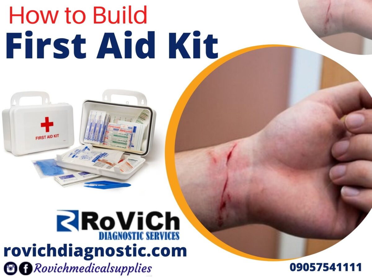
It is imperative that a well-stocked first aid kit is a must-have for treating minor injuries at home and on the go. You can buy a first aid kit or put one together on your own. Keep your supplies in a sturdy, clear plastic box so you can see what’s inside.

Rovich Diagnostics

What should be the content of the first aid kit?

How often should first aid kit be check?

Items can run out if you use them often, and medicines can expire if you rarely need them, so go through everything in your kit, and replace any empty or out-of-date items at least once a year.

Rovich Medical supplies

How and where to store first aid kit?

You don’t want to go searching for supplies when there’s a minor accident to attend to. Your kit should be easy to find. But it should be kept in a high, childproof cabinet, far away from kids’ prying fingers. Keep one full-sized kit in a central spot at home, such as your kitchen or bathroom. Then put a smaller kit in your car or purse for when you’re on the road.

How to prevent scars?

When your body heals after a cut, scrape, or burn, sometimes a scar can be left behind. Depending on the injury, some scars are small, and others are bigger and more noticeable.

To prevent scars, follow these tips:

  • Wear helmets, kneepads, and other protective gear to avoid injury.
  • Treat any cuts or other wounds right away.
  • Keep the wound moist (try an antibiotic ointment or petroleum jelly) while it heals.
  • Don’t pick at the scab.
  • Consider covering your cut with silicone gel sheeting, a clear, sticky pad that can speed healing.
  • If the scar isn’t fading, ask your doctor about creams or ointments to make it less obvious.

Hope this was helpful, for more updates contact us Rovich Diagnostics Services
first aid kit


Blue-Controller-Faux-OrganicAbstract-Technology-Gaming-Facebook-Shops-Cover-20-1200x900.jpg
31/Aug/2021

Eye Treatment

eye clinic

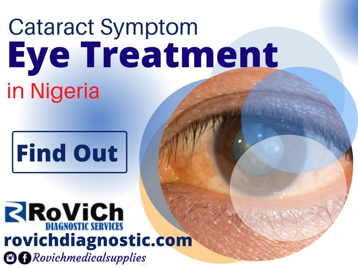
Cataract Symptom: Glare Eye Treatment

Eye Treatment

Glare, or sensitivity to light, is another symptom of cataracts. It can be difficult for a person with cataracts to see in bright sunlight. Indoor lights may begin to seem too bright, or they may appear to have halos around them. Glare from oncoming headlights can cause problems with driving at night.

Cataract Symptom: Double Vision

Double Vision

Diplopia, or double vision, when looking with one eye can be another symptom of cataracts. This is not the same as diplopia that arises from improper alignment of your eyes. The double vision seen with cataracts occurs even when you look through only one eye.

Cataract Symptom: Color Changes

eye treatment

Cataracts also affect color vision. Some colors may appear faded, and things may acquire a yellowish or brownish tint. This may not be noticed at first, but with time, distinguishing between blues and purples can be difficult.

Cataract Symptom: Second Sight

Rovich Diagnostics services

The phenomenon known as “second sight” is another characteristic of cataracts. In this situation, the cataract acts as a stronger lens, temporarily improving the ability to see things at a close distance. People who formerly needed reading glasses may no longer need them. However, as the cataract worsens over time, this temporary improvement in near vision disappears.

Cataract Symptom: New Prescription (Eye Treatment)

eye treatment

People with cataracts often need frequent changes in their eyeglasses or contact lenses because their vision deteriorates over time.

Who Can Get Cataracts?

Rovich medicals

Most cataracts occur in older people and are related to the aging process. Over half of Americans over 65 have cataracts. Sometimes, babies can be born with cataracts, known as congenital cataracts. Uncommonly, children can get cataracts as a result of illness or trauma to the eye.

Rovich Diagnostics services

What Are the Causes of Cataracts?

It is not precisely understood why people get cataracts. Aging is a known risk factor. Other factors that may also play a role in the development of cataracts include:

  • Smoking
  • Excessive use of alcohol
  • Diabetes
  • Trauma to the eyes
  • Extended use of corticosteroids
  • Prolonged radiation or sun exposure

How Are Cataracts Diagnosed?

Cataracts can be diagnosed with an eye exam. The eye exam contains a vision test and an examination of your eyes using a slit lamp microscope. The pupils are dilated with special eyedrops to provide a better view of the back of your eye, where the retina and optic nerve are located.

Cataract Surgery Eye Treatment

Surgery to remove cataracts may be required if the related vision loss cannot be corrected with glasses or contact lenses. Cataract surgery involves removing the cloudy natural lens and replacing it with an artificial lens. The operation is usually done on an outpatient basis and is very safe and effective. For those who need surgery on both eyes, the surgery is usually done on one eye at a time.

Types of Cataract Surgery

The most common type of cataract surgery is known as phacoemulsification (phaco). In this procedure, the doctor makes a tiny incision in the eye and breaks up the lens using ultrasound waves. The lens is then taken out and replaced with an intraocular lens (IOL).

Another type of cataract surgery is called extracapsular cataract surgery. This procedure involves a larger incision and removal of the cloudy lens in one piece. In most cases, placement of an IOL eliminates the need for thick eyeglasses or contact lenses.

Innovations In Cataract Surgery (Eye Treatment)

New developments in cataract surgery allow for procedures that correct both near and distance vision, reducing or even eliminating the need for glasses after the operation. Conventional “monofocal” lenses only correct distance vision, so people still need reading glasses after surgery. So-called “toric” implants are available to correct astigmatism. This picture illustrates a lens in development (shown next to a dime) that offers better color vision.

What to Expect After Cataract Surgery

After surgery, your eyes may itch and feel sensitive to light for a few days. You may need to wear a shield or glasses for protection, and you may be prescribed eyedrops to speed the healing process (Eye Treatment). It takes about 8 weeks for the eye to completely heal even though changes in vision are apparent shortly following the surgery. You may still need glasses for distance vision or reading, after the surgery, and it is likely that you will require a new prescription after your eye has healed.

Hope this was helpful, for more update involving eye contact us

Eye Treatment


Blue-Controller-Faux-OrganicAbstract-Technology-Gaming-Facebook-Shops-Cover-18-1200x900.jpg
30/Aug/2021

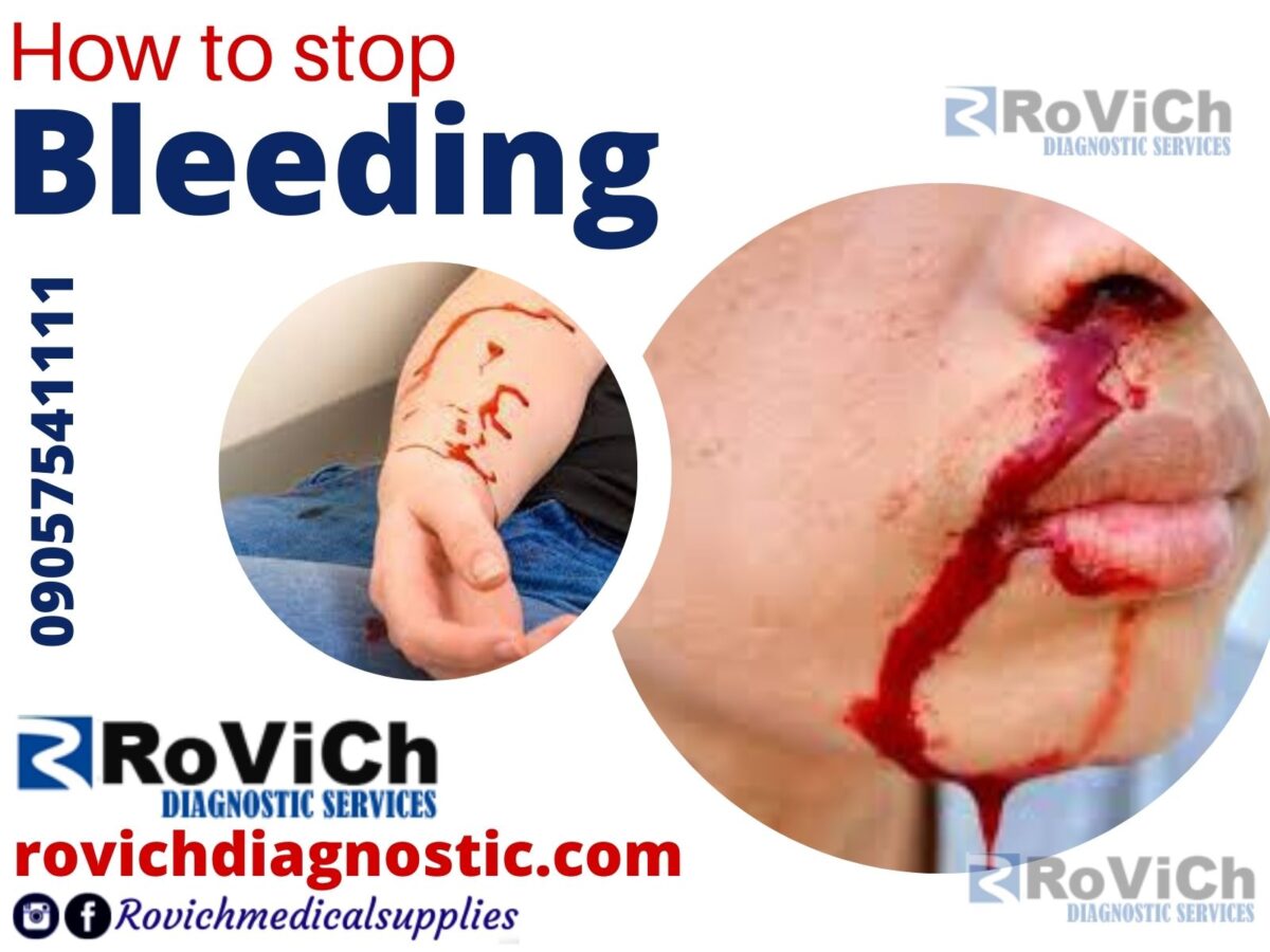
How to stop Bleeding


INTRODUCTION HOW TO STOP BLEEDING

Bleeding time is a medical test done on someone to assess their platelets function. It involves making a patient bleed then timing how long it takes for them to stop bleeding. The term template bleeding time is used when the test is performed to standardized parameters. The bleeding time test is a method indicated when other more reliable and less invasive tests for determining coagulation are not available. However, it remains the most reliable way of assessing clinical bleeding in patients with uremia.Historically it was indicated whenever the physician needed information about platelet activation. The process involves cutting the underside of the subject’s forearm, in an area where there is no hair or visible veins. The cut is of a standardized width and depth, and is done quickly by an automatic device. A blood pressure cuff is used above the wound, to maintain venous pressure at a specified value. The time it takes for the bleeding to stop (i.e. the time it takes for a platelet plug to form) is measured. Cessation of bleeding can be determined by blotting away the blood every several seconds until the site looks “glassy”. Bleeding time is affected by platelet function, certain vascular disorders and von Willebrand Disease—not by other coagulation factors such as haemophilia. Diseases that cause prolonged bleeding time include thrombocytopenia, disseminated intravascular coagulation (DIC), Bernard-Soulier disease, and Glanzmann’s thrombasthenia. Aspirin and other cyclooxygenase inhibitors can significantly prolong bleeding time. While warfarin and heparin have their major effects on coagulation factors, an increased bleeding time is sometimes seen with use of these medications as well. People with von Willebrand disease usually experience increased bleeding time, as von Willebrand factor is a platelet adhesion protein, but this is not considered an effective diagnostic test for this condition. It is also prolonged in hypofibrinogenemia. 

Medical Consultation

How to test for bleeding time

A blood pressure cuff is inflated around your upper arm. While the cuff is on your arm, the health care provider makes two small cuts on the lower arm. They are just deep enough to cause a tiny amount of bleeding. The blood pressure cuff is immediately deflated. Blotting paper is touched to the cuts every 30 seconds until the bleeding stops. The provider records the time it takes for the cuts to stop bleeding. A standard-sized incision is made around 10 mm long and 1 mm deep. The time from when the incision is made until all bleeding has stopped is measured and is called the bleeding time. Every 30 seconds, filter paper or a paper towel is used to draw off the blood.

A forecast result :  Bleeding normally stops within 1 to 9 minutes. However, values may vary from lab to lab. A prolonged bleeding time may be a result from decreased number of thrombocytes [platelets] or impaired blood vessels. However, the depth of the puncture or incision may be the source of error.

rovich diagnostics  

How to stop Bleeding 2021 Review

Bleeding time is a medical test that measures how fast small blood vessels in the skin stop bleeding. The bleeding time test is used to evaluate how well a person’s blood is clotting. The test evaluates how long it takes the vessels cut to constrict and how long it takes for platelets in the blood to seal off the hole. where the pre-operative assessment of patients taking aspirin or NSAIDs and screening for von-Willebrand disease.

 NOTE :  A prolonged bleeding time may be a result from decreased number of thrombocytes [platelets] or impaired blood vessels

Diseases that cause prolonged bleeding time include

  • von Willebrand disease — a genetic disorder caused by a missing or defective clotting protein
  • thrombocytopenia — a deficiency of blood platelets
  • disseminated intravascular coagulation(DIC) — widespread formation of blood clots in the small blood vessels throughout the body
  • Glanzmann’s thrombasthenia — a genetic disorder in which the platelets have a deficient fibrinogen receptor
  • hypofibrinogenemia — a partial deficiency of fibrinogen


Frequently ask question 

What are the 3 stages of blood clotting?

  •  Constriction of the blood vessel
  •  Formation of a temporary “platelet plug.
  •  Activation of the coagulation cascade
  •  Formation of “fibrin plug” or the final clot.

How to stop Bleeding

What is the risk involved during the bleeding time test

There is a very slight risk of infection where the skin is cut.

What are the factors affecting bleeding time?

The bleeding time is affected by the antiplatelet variables of

 all of which interfere with accurate interpretation of the test results.

CONCLUSION

Most people will never need a bleeding time test. You may need to have a bleeding test if you’ve been experiencing bleeding that won’t stop, especially from small incisions, punctures, or cuts. Your doctor can choose from a number of tests to evaluate your platelet function. A bleeding time test is a common test to screen patients having prolonged bleeding times. Abnormal results from a bleeding time test can be a sign that you need more in-depth testing to find the cause of your prolonged bleeding. It could mean you have an acquired platelet function defect, which is a condition that develops after birth and affects how well your blood platelets work. Your body may produce too many or too few platelets, or your platelets may not work properly.

Hope this was helpful for more information contact us

How to stop Bleeding


Private-over-Federal-Eye-clinic-1200x900.jpg
27/Aug/2021

Eye clinic

rovich diagnostic services

One of The greatest gifts of God to man is the gift of vision, every activity of man is by the power of vision, and when it is lost it seems all hope and dreams are gone. Therefore taking proper care of the eye should be a priority for everyone since it cannot be replaced. The medical sector has shown great ability to explore the study of the human body system which has proven successful through the help of technology as it has with the outcome of optometry.

In this context, I will talk about federal clinics and private eye clinics in Nigeria with a brief history as it will help identify the best medical eye clinic to maintain a clear vision for as long as we live. Give yourself the gift of clear vision – schedule an appointment with your eye care provider today.

 What is clinic 

A clinic sometimes refers to as a specialist hospital is a healthcare facility primarily focused on the care of outpatients who need special attention on their health issues. Clinics can be privately operated or publicly managed and funded. They typically cover the primary care needs of populations in local communities, in contrast to larger hospitals that offer more specialized treatments and admit patients for overnight stays.

The word clinic refers to a general practice, run by one or more medical practitioners offering small treatments, but it can also mean a specialist clinic. Family Eye Clinic has been a leading provider of optometry services and vision care products since 1995 and has proven to help you achieve and maintain a clear vision for years to come.

eye clinic

Federal eye clinic

Visual impairment and blindness due to ocular diseases are significant public health problems in many parts of the world. According to the World Health Organization on Report in 2010, about 285 million people are visually impaired worldwide and out of these numbers, 39 million (14%) are blind while 246 million (86%) have low vision or severe visual impairment and 90% of them live in developing countries with a poor economy. Nigeria with an estimated population of about 200 million people (National Bureau of Statistics, 2012) is the most populated country in Africa. The results of the blindness and low vision survey conducted from 2005 – 2007 reported the prevalence of blindness and visual impairment at 0.78% for all ages and 4.2% for people over 40 years. If eye care services are made accessible to people, the prevalence of avoidable causes of blindness will be reduced. According to Nigeria’s national visual impairment and blindness survey, the northwest geopolitical zone has the highest number of blind people (28.6%). Both urban and rural dwellers in the Federal Capital Territory exhibit similarity in the prevalence of eye diseases yet there is a significant difference in their utilization of eye care services.

Cases the federal eye clinic can handle include 

  • Age-related macular degeneration
  • Blind eye removal and rehabilitative socket surgery
  • Cataracts eye problem 
  • Corneal disease and conditions
  • Diabetic eye disease
  • Diabetic retinopathy
  • Eyelid cancers
  • Glaucoma
  • Lens implants for visual correction
  • Macular hole
  • Oculoplastics for eyelid malpositions, lumps, tumors, and cysts
  • Pediatric eye problems
  • Squint
  • Uveitis
  • Watery eyes and blocked tear ducts
  • & much more.

eye clinic

ADVANTAGES OF FEDERAL EYE CLINIC

  • Free media eye examinations are done before treatment
  • All eye specialists working in the clinic are fully employed.
  • Eyeglass repairs are done free without payment.
  • The federal clinic has up to three or more theaters where eye surgery are been carried out.
  • Charges for operation are very low compare to private.
  • There is a chance of getting a quick response for operation.


DISADVANTAGES OF FEDERAL EYE CLINIC 

  • There is no privacy in the clinic
  • There is always a large number of people waiting to see the specialist 
  • A proper medical checkup is not giving.
  • The attention of a patient to a specialist is very low.


FACTORS THAT AFFECT UTILIZATION OF EYE CARE SERVICES  

Studies within and outside Nigeria have implicated gender, cost, ignorance, attitude, lack of education, and urban residence as common reasons that contribute to the utilization of eye care services. In Nigeria and the Federal Capital Territory (FCT), eye care services are provided by ophthalmologists, optometrists, ophthalmic nurses, and dispensing opticians. The available services range from routine eye examinations to invasive surgical procedures. The majority of the eye facilitates are located in the cities and operational hours are usually from 8.00 a.m to 6.00 p.m on weekdays and only a few operate on weekends. The consequence is the mal-distribution of skilled personnel and facilities. Evidence suggests that improved access to adequate health care leads to enhanced health outcomes. If eye care services are accessible, people who need the services will use them. The prevalence of blindness and visual impairment is high in Nigeria, determining the barriers to access eye care services is essential for developing strategies to prevent blindness.

Exercise

PRIVATE EYE CLINIC

A private clinic is any healthcare building where private practice is carried out on outpatient basis and observation purposes before referral or transfer to a hospital for further management.

The private eye clinic runs ophthalmology which is a branch of medicine dealing with the diagnosis, treatment, and prevention of eye and vision diseases. The Private Hospital offers outpatient clinics with consultant ophthalmologists every weekday in a dedicated ophthalmic suite. Should you require further treatment, the private eye clinic has a theatre where surgery can be carried out. It is little or no waiting time for surgery and treatment can start as soon as patients are ready. Experienced consultant ophthalmologists are all experts at diagnosing and treating eye conditions.


It might interest you to know that the most common operations done in private clinics include one-day cataract surgery and hip and knee replacements. Some of these procedures typically take weeks to recover from and not surprisingly, many patients would prefer to recover from the comfort of their homes.


Cases the private clinic can handle involve 

  • Cataracts
  • Diabetic retinopathy
  • Glaucoma
  • Lens implants for visual correction
  • Macular hole
  • Pediatric eye problems
  • Watery eyes and blocked tear ducts
  • Pregnancy operations.


ADVANTAGES OF PRIVATE CLINIC 

  • Private clinics provide patients with privacy.
  • Excellent doctor-to-patient ratios.
  • State-of-the-art medical equipment.
  • Upscale amenities.
  • Faster access to the best medical specialists.


DISADVANTAGES OF PRIVATE CLINICS

  • Specialists are consultants, not employers
  • Expenses are very high 
  • A late response to emergency operation 
  • There is no free eye check-up
  • On free medical eyeglasses for patient 
  • Has only a small theater for an eye operation

Types of Ultrasound

Privately operated clinics have their pros and cons, These clinics are completely legal as long as they are following industry standards and comply with the Ministry’s billing laws. Despite the cost, they are becoming more popular than ever. When you consider the higher level of personal attention and the convenience, the higher cost of private clinics doesn’t sound so bad to some people. Even though private clinics often don’t take health insurance, paying the money out-of-pocket can have its benefits. Being able to skip the waiting line and have your procedure done by the doctor of your choice within days is a great incentive. Private clinics can be found across the country, a simple Google search can help you find one close to you that suits your individual needs.


FREQUENTLY ASKED QUESTION

 QUESTION 1

Are private clinics better than public?

Most federal clinics are better than private clinics as they have more medical eye facilities for any eye condition and are usually equipped to handle more complex cases.


QUESTION 2

What are the symptoms of eye problems?

Answer 

There are different types of eye problems with different symptoms, the most common eye problems symptoms are long-sightedness and short-sightedness, poor vision, itching of the eyes, water running eyes. Good healthy eyes can see objects from far and near.  

Question 3

What can damage the eye lens?

Answer 

The eye lens is very sensitive and can be damaged by direct contact with sharp objects, direct exposure to sunlight or high-intensity light rays, and exposure to chemicals. 


Question 4

Who should I consult for eye treatment?

Answer 

There are doctors that specialize in eye treatment and they are the best to handle any eye problems. They are mostly found in federal medical clinics (FMC) and are known as opticians. 


Question 5.

Where can I get good medical eyecare facilities in Nigeria? 

Answer 

The best place to get all medical technology with good quality is at Rovich Diagnostics Services.


Conclusion 

As waiting months for a procedure can be both extremely inconvenient and uncomfortable, private practices can provide a quicker alternative. While many surgeries in the hospital are covered by insurance, surgeons can charge hundreds or even thousands of dollars for those exact same procedures in their own clinics. Although more respondents were aware of the need for a regular eye check, the utilization was low. The study has identified no felt need as the major reason why people in the Bwari area council do not access eye care services. The cost was also a factor. The other reason why individuals were not accessing eye care services was the non-availability of eye clinics/centers. More effort needs to be made to get more people to utilize the eye clinic. These include awareness and health education campaigns on the need for a regular eye check-up. Development of strategies for accessible and affordable eye care services.

eye clinic


Blue-Controller-Faux-OrganicAbstract-Technology-Gaming-Facebook-Shops-Cover-12-1200x900.jpg
23/Aug/2021

Federal Hospital

Federal Hospital in Nigeria. Since no one wants to risk his or her life and take a risk in the slightest negligence in treatment, we all have a choice to choose what is best for us to stay alive, but we often forget to asked ourselves if these(snacks, drink, meal, way of life) are healthy for us? This question strikes us to caution our actions for better healthy living as the hospital is no place one could wish to be. An old phrase would say “You never miss your health until you lose it”. health shouldn’t be taken for granted as it is the real wealth and not pieces of gold and silver. I understand for one to have a healthy body you need to have (exercise, good thoughts, eating good food, taking a good sleep)  which tends to be highly repetitive when engaged in it regularly, as it is a major driver of neuroplastic changes in the brain and body system, which is why physical exercise is one of the most important factors in restoring and maintaining mental as well as physical health across the life span, starting from the moment one begins such a program.

Generally, hospitals have evolved since the 1700s when the first U.S. public hospital sheltered and provided medical healthcare to the poor. Until the late 20th century, public hospitals represented the “poor house” that undertook social welfare roles. The “poor house” also provided secondary medical care, specifically during epidemics. Let’s focus on medical centers where treatments of different illnesses can best be handled by experts in the field to restore and maintain good healthcare in Nigeria. There are factors to consider when choosing a hospital for medication. For non-emergency situations – such as childbirth planning or undergoing scheduled or routine medical procedures – patients often have a choice of what type of hospital to go to, either public or private.

Best medical consultation

Introduction 

A hospital is a health care institution providing patients treatment with specialized medical and nursing staff with full medical equipment or facilities. Specialized hospitals can help reduce health care costs compared to general hospitals.is because the specialist practitioner has all the medical facilities to handle any health problems more effectively as it relates to its line of specialization. The hospital which is best known to have an emergency department to treat urgent health problems is the general hospital, caring for problems ranging from fire and accident victims, sudden illness leading to deformation or death of the patient. With so many different types of hospitals and medical facilities, you may wonder what makes one hospital or facility different from another, with a good medical practitioner and facilities a whole lot of difference can be made. Specialized hospitals include trauma centers, rehabilitation hospitals, children’s hospitals, seniors’ (geriatric) hospitals, and hospitals for dealing with specific medical needs such as psychiatric treatment and certain disease categories. Hospitals are classified as general, specialty, or government depending on the sources of income received.

The Federal hospital

Firstly, with the anticipation of free health services, there is a high likelihood of poor quality of services. The sole responsibility of any government is to provide basic amenities for its citizens. One of the key areas governments focus their attention on is public health care which renders public services to every citizen of the state. Federal hospitals do have benefits as they are equipped with the latest technology and healthcare materials, as Most federal hospitals have strict rules and policies when it comes to treating people. Citizens have the benefit of receiving free health care services through a federal health care center.

Federal Hospital

A federal hospital some times refer to as a public hospital is any medical healthcare center owned by the government of the people and is fully funded by the government and operates solely off the money that is collected from taxpayers to fund healthcare initiatives thereby creating employment for doctors and specialist in a different field. In some countries, this type of hospital provides medical care free of charge to patients, covering expenses and wages by government reimbursement. Although the services provided by public and private hospitals have some similarities with a little difference. Since costs tend to be lower in publicly operated hospitals, it is the best option for those who have restrictive insurance or who are not wealthy and able to pay for their healthcare out of pocket.

Medical System

ADVANTAGES OF FEDERAL HOSPITAL

  • Federal or Public hospitals are much larger than private hospitals. It has a different ward for different illnesses such as maternity ward for children, men, pregnant women and people with disabilities, etc.
  • Federal hospitals provide the best and latest technology and healthcare materials needed to treat and diagnose illnesses. 
  • Public hospitals are funded by the government and so are unable to turn away patients with any kind of illness or emergency.
  • The cost of treatment is considerably cheaper than a state hospital and they have a resident doctor in case of an emergency.
  • The federal hospital is owned and controlled by the federal government and is fully funded by the Government which therefore creates more employment for medical practitioners and training of aspiring doctors and nurses.
  • Because they are partly or fully funded by the federal government, federal hospitals accept nearly every type of insurance and are very flexible.
  • They also have a much higher number of beds, so they can accommodate more patients at a time.
  • The federal hospital has a good standard of medical facilities required to handle any emergency.


THE DISADVANTAGES OF FEDERAL HOSPITAL 

  • Due to the influx of people which leads to overcrowding, one may have trouble obtaining medical care as it will prolong or delay medical attention.
  • In the federal hospital doctors and nurses are not always available especially if a patient requires immediate treatment or has a suspected serious illness.
  • There is no privacy in the federal hospital which one is prone to contacting another illness from the environment.
  • Cheaper and less effective medication may be given in place of quality and effective drugs. It should not be expected that citizens have the best quality of health services due to the limited budget and expenditure of the ministry. 


THE STATE HOSPITAL

With the poor performance of the federal government’s low budget in providing good medical care for citizens in all states, the state government took it upon itself to assist with the best medical facilities needed to treat and handle any medical illnesses. This comes with a higher maintenance cost as most of the state cant afford a well-standard fully equipped hospital for her citizens. You will see that people tend to visit private hospitals more than public hospitals.

A state hospital is any medical healthcare establishment that is licensed by the ministry of health to provide medical attention to citizens under medical need, as it is owned and funded by the state government without the support of federal fund thereof all operational medical activities and payment of staffs are run by the state government, for-profits and non-profits making. State hospitals are commonly part of the health development in any country as they will help to save lives.


ADVANTAGES OF STATE HOSPITAL

  • State hospitals have a short period of waiting time.
  • A state hospital enables the patient to select their preferred surgeon or doctor for treatment.
  • State hospitals offer an excellent doctor-to-patient ratio. In addition, there is usually a larger number of attentive staff (nurses) per patient in state hospitals.


DISADVANTAGES OF STATE HOSPITAL

  • State hospitals are not easily affordable as services in these settings tend to be much higher.
  • State hospitals can refuse treatment due to lack of healthcare equipment, as they might refer you to a private or federal hospital.
  • State or local hospitals are not well secured as a result of poor employment of security guards.
  • They are unhygienic as cleaners are not employed as much to keep the environment clean.
  • They do not conduct proper medical checkups and lack drugs in the hospital pharmacy.
  • The doctors are not very well educated to handle surgical analysis as they don’t have a resident doctor.
  • In the state or local hospital there is a lack of proper communication.
  • They have no ambulance for emergencies.

Federal Hospital

FREQUENTLY ASKED QUESTIONS

QUESTION 1

 What is the difference between a teaching hospital and a maternity hospital?

Answer 

A teaching hospital is a healthcare hospital that has an accredited medical residency-training program and usually has an affiliation with a medical school to train students or persons aspiring to be a nurse in the medical practice

While

Maternity hospital specializes in caring for women during pregnancy and childbirth. It also provides care for newborn infants and may act as a center for clinical training in midwifery and obstetrics. 

QUESTION 2.

Can I treat malaria by taking antibiotics every month?

Answer 

Firstly, taking drugs without running a medical test and prescription by a doctor or pharmacist is drug abuse. Antibiotics are not healthy when taken often and are not advisable. 

QUESTION 3.

How can we improve a state hospital?

Answer 

Improving a state hospital requires the state government to

  1. Employ qualify doctors and nurses 
  2. Purchase new technologies for medical treatments of patients
  3. Provide state pharmacists with original drugs
  4. Employ cleaners to keep the environment clean
  5. Provide a complete laboratory and ultra-scan machine for diagnosis of illness.

It is very easy to purchase all kinds of modern medical facilities in Nigeria, at Rovich Diagnostics Services we provide all kinds of technology for the medical sections and all engineering sections. Our services are the best for any changes in your business. 

CONCLUSION 

Choosing the right hospital and physician are important factors to consider that significantly influence a patient’s treatment. The preferred choice for many patients in choosing a hospital is dependent on the location and type of illness. Rovich Diagnostics Services  got you covered in all kinds of physiotherapy, Eye clinical issues and many more.

Even though hospitals are mostly funded by taxpayers, some hospitals, as well as medical research facilities, receive charitable donations. Besides this, there is an increasing trend of privatization of some hospital services if those services go beyond provincial health budgets. All hospital federal and state hospitals should be well taken care of as the health of the citizens is a priority for the government.


rovich diagnostic services


Blue-Controller-Faux-OrganicAbstract-Technology-Gaming-Facebook-Shops-Cover-3-1200x900.png
20/Aug/2021

rovich diagnostic services

Eye problem in Nigeria

Nearsightedness (Myopia)

Eye problem in Nigeria

Eye problem in Nigeria. It is said Nearsightedness (myopia) causes people to be unable to see distant objects, though they can see nearby objects clearly. It is caused by the cornea having too much curvature, resulting in problems with focusing on the retina. Myopia is extremely common and easily corrected with eyeglasses, contact lenses, or surgery.

Eye problem in Nigeria Color Blindness

Eye problem in Nigeria

It has been proven that the colors we see are a result of how our eyes (and thus our brains) interpret different wavelengths of light. People with color blindness have difficulty seeing certain colors, usually reds, greens, and blues. Color blindness is caused by an absence or malfunction of color-sensitive cells located in the retina. Most of the time this is genetic (people are born with it) but it can also be caused by aging, disease, trauma to the eye, or certain medications. If the cause of the color blindness is genetic, the problem cannot be corrected but people may be trained to adapt to interpret color shades. In cases where color blindness is acquired, it may be treatable.

Eye problem in Nigeria Cataract Symptom: Blurry Vision

Eye problem in Nigeria

The major symptom of cataracts is seeing blurry images at any distance. People may describe their vision as foggy, cloudy, or filmy. Cataracts get worse with time, and less light reaches the retina. It may be especially hard for people with cataracts to see and drive at night.

Eye problem in Nigeria Astigmatism

Eye problem in Nigeria

Astigmatism is another another cause of visual difficulty, in which images are blurred due to an irregularly-shaped cornea. Astigmatism will eventually affect most people as a part of the aging process. It is treated with glasses, contact lenses, or refractive laser eye surgery.

Eye problem in Nigeria

Proactive Eye Health

Dealing with Eye problem in Nigeria. It is important to take care of your eyes to protect your vision. Always use eye protection to avoid injuries and wear sunglasses to protect your eyes from UV rays. People over 40 should have their eyes checked every two years and people over 60 should have their eyes checked every year.

Be proactive in your eye health. If you notice any symptoms of the disorders discussed in this slide show, see your ophthalmologist.

What Could these Cataracts be?

Studies has shown that Cataracts are a painless clouding of the internal lens of your eye. Because they block light from passing through the lens, cataracts make it difficult to see clearly and can even scause blindnes over time. Cataracts are progressive, meaning they worsen with time. Most cases occur in older people, but sometimes they can be seen in younger people as well.

How Your Vision Is Affected By Cataracts

Light enters your eye and passes through the lens. The lens of your eye focuses light onto the retina, which transmits visual signals through the optic nerve to your brain. Clouding of the lens due to cataracts results in blurring of the images you see. Other problems with the eyes can also cause blurry vision, but cataracts produce some characteristic symptoms.

Hope this was helpful, on Eye problem in Nigeria. Follow us for more info and updates Rovich Diagnostics Services

rovich diagnostic services


Blue-Controller-Faux-OrganicAbstract-Technology-Gaming-Facebook-Shops-Cover-9-1200x900.jpg
18/Aug/2021

Types of Ultrasound scan

An ultrasound scan is a medical test that uses high-frequency sound waves to capture live images from the inside of your body. It’s also known as sonography. The technology is similar to that used by sonar and radar, which help the military detect planes and ships. Types of Ultrasound allows your doctor to see problems with organs, vessels, and tissues without needing to make an incision. Unlike other imaging techniques, ultrasound uses no radiation. For this reason, it’s the preferred method for viewing a developing fetus during pregnancy. Also Ultrasound or ultrasonography is a medical imaging technique that uses high frequency sound waves and their echoes. The technique is similar to the echolocation used by bats, whales and dolphins, as well as SONAR used by submarines. Ultrasound was originally developed during World War I to track submarines as SONAR technology (Sound, Navigation And Ranging). Ultrasound was first used medically in the 1950s, with very early applications in fetal biometry; nowadays, it is used in just about every field of medicine. Furthermore, it is also now practiced by a wide variety of professionals, in a multidisciplinary setting. The Types of Ultrasound listed below is a high frequency sound beyond the hearing of the human ear. The frequencies of ultrasound required for diagnostic medical imaging are in the range 1–20 MHz. These frequencies can be obtained by using piezoelectric materials (particularly crystals). When an electric current is applied and reversed across a slice of one of these materials, the material contracts or expands. So a rapidly alternating electric field can cause a crystal to vibrate. These vibrations are then passed through any adjacent materials, or into the air as a longitudinal wave is produced – a sound wave


Types of Ultrasounds 

Abdominal Ultrasound

An abdominal ultrasound is a useful way of examining internal organs, including the liver, gallbladder, spleen, pancreas, kidneys, and bladder. This can help to diagnose a variety of conditions and to assess the damage caused by illness. Because it provides real-time images, ultrasound can also be used to:

  • Guide procedures such as needle biopsies, in which needles are used to sample cells from organs for laboratory testing.
  • Help a physician determine the source of many abdominal pains, such as stones in the gallbladder or kidney.
  • Help identify the cause for enlargement of an abdominal organ.
  • Doppler ultrasound is a special type of ultrasound study that examines major blood vessels. These images can help the physician to see and evaluate:
  • Blockages to blood flow, such as clots.
  • Build-up of plaque inside the vessel.
  • Congenital malformation.
  • Types of Ultrasound scan

Pelvic Ultrasound Imaging

Pelvic ultrasounds are one of the most well-known forms of ultrasound, being one of the imaging tests used to monitor the health of the embryo or fetus during pregnancy. Aside from maternity medicine, these ultrasounds are also used to examine the uterus, ovaries, bladder, and prostate gland. Pelvic ultrasound is often used to diagnose conditions or the cause of conditions such as:

  • Pelvic pain
  • Abnormal bleeding
  • Menstrual problems
  • Ovarian cysts
  • Uterine fibroids
  • Ovarian and uterine cancers
  • Kidney and bladder stones

Transabdominal

Patients receiving a transabdominal ultrasound need to have a full urinary bladder. Like other ultrasound procedures, patients lie on their back as a gel is applied to the abdomen. The transducer is then rubbed over the examination area and releases sound waves. This is a fairly straightforward ultrasound exam.

Transvaginal Ultrasound

For a transvaginal ultrasound, a woman needs to empty her bladder the same way she would for a gynecological exam. She also lies face up on her back with feet in stirrups. The transducer of the ultrasound needs to be inserted for this test. The transducer is smaller than the standard speculum used in Pap tests. A protective cover and gel for lubrication is placed on the transducer before it is inserted in the vagina.

Only the first two to three inches of the transducer is inserted in the vagina. The doctor may move it around to obtain images from different angles. The most common reason for transvaginal pelvic ultrasounds is to look for the cause of pelvic pain. Most patients report that this exam is more comfortable than a manual gynecologic examination.

Types of Ultrasound scan

Transrectal

In order to perform an ultrasound on the prostate gland, the transducer must be inserted through the rectum so that the sound waves can travel to the prostate. As with other inserted ultrasound procedures, the transducer is covered with a protective cover and lubrication before insertion. The transducer will need to be moved around in order to obtain images from different angles. These exams are typically performed with the patient lying down on their left side and knees bent up towards the chest.

The doctor may recommend a biopsy be performed if a lesion is found during the exam. In a biopsy, the radiologist uses the ultrasound images to guide a needle towards the prostate gland and extract a sample of the abnormal tissue. Ultrasound-guided biopsies are minimally invasive and only require a small incision.

Obstetric Ultrasound Imaging

Obstetric ultrasound (OB ultrasound) refers to the specialized use of sound waves to visualize and thus determine the condition of a pregnant woman and her embryo or fetus. Obstetric ultrasound should be performed only when clinically indicated. Some indications may be:

  • To establish the presence of a living embryo/fetus
  • To estimate the age of the pregnancy
  • To diagnose congenital abnormalities
  • To evaluate the position of the fetus
  • To evaluate the position of the placenta
  • To determine if there are multiple pregnancies

You will be asked to lie on your back or side. You will also be asked to expose your lower abdominal area. The obstetric ultrasound examination takes about 30-45 minutes.

This is a painless procedure. There may be varying degrees of discomfort from pressure as the sonographer guides the transducer over your abdomen, especially if you are required to have a full bladder. At times the sonographer may have to press more firmly to get closer to the embryo or fetus to better visualize the structure. This discomfort is temporary. Also, you may dislike the feeling of the water-soluble gel applied to your abdomen. With transvaginal scanning, there may be minimal discomfort as the transducer is moved in the vagina.

Best clinic for Eye

Carotid & Abdominal Aorta Ultrasound Imaging

Ultrasound of the carotid arterial system provides a fast, noninvasive means of identifying blockages of blood flow in the neck arteries to the brain that might produce a stroke or mini-stroke. Ultrasound of the abdominal aorta is primarily used to evaluate for an aneurysm which is an abnormal enlargement of the aorta usually from atherosclerotic disease.

The patient is positioned on an examination table that can tilt and move. A clear gel is applied to the area that will be examined. The gel helps the transducer make a secure contact and eliminates air pockets between the transducer and the skin, since the sound waves cannot penetrate air. The sonographer or radiologist then presses the transducer firmly against the skin and sweeps along the area of interest, reviewing the images on the monitor and capturing “snapshots” as required.

Liver Ultrasound

Liver Ultrasound determines the size, shape, and function of the liver, and can be used to detect tumors.

Renal Ultrasound

Renal ultrasound determines the size, shape, and function of the kidneys, and can be useful in the detection of kidney stones, cysts, and tumors.

Vascular Ultrasound

Vascular ultrasounds are used to analyze the flow of blood through the arteries and veins.

Thyroid Ultrasound

Thyroid Ultrasound checks for underactive and overactive thyroid glands, nodules, and cysts

Importance of ultrasound

There are multiple diagnostic uses of ultrasound. Until ultrasound came along, doctors could only listen to the foetal heart, on which presence was assumed to be an indicator of foetal well-being. With the advent of ultrasound, the information available to the pregnant patients’ attending doctors has increased markedly. Doctors are able measure the size of the foetus, which helps in determining when delivery of the baby would be expected; monitor growth of the developing foetus; check for structural abnormalities of foetus, e.g. head, and spine between 18 and 20 weeks of pregnancy; locate the site of the placenta; check for evidence of foetal well being or compromise with examinations of the foetal heart and even perform procedures on the foetus. The detection and assessment of growths in the female reproductive tract like fibroids, tubal or ovarian masses, cancers etc is another use of ultrasound. Ultrasound is used in the detection and assessment of conditions in the heart. It is used to examine the size, shape, and movement of the heart, including its valves and chambers as well as the blood flow through the heart. This type of ultrasound is called an echocardiogram, which can even be used to diagnose cardiac abnormalities in foetuses prior to their birth (foetal echocardiography). Ultrasound is used in the diagnosis of deep vein thrombosis (DVT), which is a condition in which there is blood clot formation (thrombi) in the deep veins. The thrombi can break loose and travel to the lungs, causing pulmonary embolism, which is a life threatening condition. Abnormal conditions in other organs that can be detected and assessed by ultrasound include the liver, gallbladder, pancreas, kidneys, bladder, prostate, testes, breasts, thyroid, lymph nodes, skin, joints, and eyes. Procedures like biopsies, in which tissue samples are taken for analysis, can be carried out under ultrasound guidance. The types of ultrasound listed below are  used as a guide to the correct location of the site for the biopsy to be carried out. Examples include breast and prostate biopsies.

Best medical consultation

Limitations

The limitations to the use of ultrasound are due to its properties, viz: ultrasound waves cannot pass through bone, air, or gas. Therefore, it is not possible to produce detailed images of some parts of the body like the brain, which is surrounded by bone. Doctors utilise other imaging methods like CT scans and magnetic resonance imaging (MRI) scans to examine parts of the body that are not suitable for ultrasound examination.

Mechanism of an ultrasound scan

  1. The ultrasound machine transmits high-frequency (1 to 5 megahertz) sound pulses into your body using a probe.
  2. The sound waves travel into your body and hit a boundary between tissues (e.g. between fluid and soft tissue, soft tissue and bone).
  3. Some of the sound waves get reflected back to the probe, while some travel on further until they reach another boundary and get reflected.
  4. The reflected waves are picked up by the probe and relayed to the machine.
  5. The machine calculates the distance from the probe to the tissue or organ (boundaries) using the speed of sound in tissue (5,005 ft/s or1,540 m/s) and the time of each echo’s return (usually on the order of millionths of a second).
  6. The machine displays the distances and intensities of the echoes on the screen, forming a two dimensional image like the one shown below.

Frequently ask question on Types of Ultrasound

What happens after the test?

A radiologist will interpret your ultrasound images. Your doctor will discuss the results with you at a follow-up appointment. Your doctor may ask for another follow-up scan or other tests and set up an appointment to check on any issues that were found.

The difference between sonogram and types of Ultrasound

Often, the terms sonogram and ultrasound are used interchangeably. However, there’s a difference between the two:

  • An ultrasound is a tool used to take a picture.
  • A sonogram is the picture that the ultrasound generates.
  • Sonography is the use of an ultrasound tool for diagnostic purposes.

NOTE : an ultrasound is the process, while a sonogram is the end result.

rovich diagnostic services

Types of Ultrasound

Sonography is a noninvasive, painless procedure. It uses high-frequency sound waves — called ultrasound waves —to produce images of organs, soft tissues, blood vessels, and blood flow, from inside the body. These images are used for medical analysis.

After x-ray exams, ultrasound is the most commonly used form of diagnostic imaging. It helps doctors gain insights into the inner workings of the body, and is known for being:

  • safe
  • radiation free
  • noninvasive
  • portable
  • widely accessible
  • affordable

Sonogram

A sonogram (also called an ultrasonogram) is the visual image produced during an ultrasound examination.

What is these Types of Ultrasound used for?

Probably best known for confirming and monitoring pregnancy, ultrasound is also commonly used by doctors for:

Diagnostics

Doctors use ultrasound imaging to help diagnose conditions affecting the organs and soft tissues of the body, including:

  • abdomen
  • liver
  • kidneys
  • heart
  • blood vessels
  • gallbladder
  • spleen
  • pancreas
  • thyroid
  • bladder
  • breast
  • ovaries
  • testicles
  • eyes

There are some diagnostic limitations for ultrasounds. For example, sound waves do not transmit well through areas that might hold gas or air (such as intestines), or areas blocked by dense bone.

Medical procedures

When a doctor needs to remove tissue from a very precise area in the body — such as in a needle biopsy — ultrasound imaging can help with visual direction.
best Medical Consultation

Conclusion of Types of Ultrasound

Historically, the diagnosis of appendicitis has been made based on clinical findings. Diagnostic imaging has been used primarily to evaluate patients who have an atypical clinical presentation. Over the past several years, improvements in imaging technology have contributed to an increase in diagnostic accuracy in these patients.

These Types of Ultrasound has been suggested and used as the primary diagnostic imaging modality to evaluate for appendicitis. However, sonography is known to be highly operator dependent; large patient habitus and atypical appendiceal location are additional factors that may reduce the reliability of a negative sonographic examination for appendicitis.

CT is more accurate than ultrasound in the diagnosis of acute appendicitis. When dedicated CT examination of the appendix is performed to evaluate all patients with clinically suspected appendicitis, the diagnostic accuracy is 98%. Early and accurate diagnosis of appendicitis can decrease patient morbidity and hospital costs by reducing the delay in diagnosis of appendicitis and its associated complications, as well as by avoiding inpatient observation prior to surgery in patients who present with atypical symptoms. Furthermore, both CT and ultrasound may rapidly provide alternative diagnoses which can be treated on an outpatient basis.

Types of Ultrasound scan

Hope this was helpful, for more updates contact Us Rovich Diagnostics Service


Blue-Controller-Faux-OrganicAbstract-Technology-Gaming-Facebook-Shops-Cover-7-1200x900.jpg
16/Aug/2021

Best clinic for Eye

Best clinic for Eye

Chalazion (Eyelid Cyst)

Chalazion (Eyelid Cyst) A chalazion (also called a meibomian cyst, tarsal cyst, or conjunctival granuloma) is the inflammation of a small cystic gland in the eyelid. The gland opening becomes clogged and the gland swells. Chalazions are treated with warm compresses, though in rare cases they may require antibiotics. If the chalazion becomes severe, causes changes in vision, or is persistent, it may be removed surgically. Corneal Ulcer A corneal ulcer is a common eye condition. It refers to small crater (ulcer) on the front part of the eye, usually following an infection. Bacteria, viruses, or fungus can cause a corneal ulcer. People who wear contact lenses are at higher risk for corneal ulcers. That’s because infectious agents may get trapped behind a lens. People deficient in vitamin A are particularly vulnerable to corneal ulcers. Symptoms of a corneal ulcer include: • pain, • intense redness, • feeling as if the eye is scratched or something is in the eye, • sensitivity to light, and • blurry vision. If you suspect a corneal ulcer or have the symptoms of a corneal ulcer and wear contact lenses, see your ophthalmologist immediately. High potency antibiotics and pain medications are the treatments for this condition. Diabetic Retinopathy People with diabetes often have problems with their blood vessels throughout their bodies and the eye is no exception. Diabetes can come with complications, such as diabetic retinopathy. This eye problem affects the blood vessels in the back of the eye, on the retina. There are two types of diabetic retinopathy: • Nonproliferative retinopathy, the less severe type in which there may be bleeding in the retina and leakage of blood or serum causing a "wet retina." • Proliferative retinopathy, a more severe type where new abnormal blood vessels grow on the retina. These vessels may bleed into the vitreous (the clear jelly in the center of the eye) and cause visual problems. Treatment involves laser surgery but damage may be permanent. The best way to prevent diabetic retinopathy is with strict glucose control and a healthy lifestyle (weight loss, dietary restrictions, and exercise). Diabetes and Dry Eyes Multiple studies have shown a relationship between diabetes and dry eyes. What’s more, studies suggest that the higher your blood sugar levels spike, the more likely you are to have dry eyes. This eye condition is mainly treated with eye drops, blocking tear ducts, or medicine that increases how much your eyes tear. Crossed Eyes (Strabismus) Crossed eyes (strabismus) is an eye problem involving a misalignment of your eyes. In other words, your eyes do not look in the same direction as they should. One eye may track differently than the other, causing a disjointed appearance. Young children born with this disorder may develop decreased vision in one eye (amblyopia, also known as “lazy eye.”). Treatment for strabismus involves using an eye patch on the stronger eye, eye exercises, and possibly surgery. Floaters Floaters are caused by aging changes in the vitreous jelly of the eye. They are a common consequence of aging. If you develop multiple floaters, or floaters associated with pain, get checked by your ophthalmologist. In general floaters do not cause blindness and are mostly harmless. There is no definitive treatment for floaters, as most will fade or become less noticeable over time. Farsightedness (Hyperopia) Farsightedness (hyperopia) is difficulty focusing on objects that are close. It is very common and the incidence increases with age. It is caused by an abnormally flat cornea that does not allow light to sharply focus on the retina. Glasses, contact lenses, or surgery may be used to correct hyperopia.A chalazion (also called a meibomian cyst, tarsal cyst, or conjunctival granuloma) is the inflammation of a small cystic gland in the eyelid. The gland opening becomes clogged and the gland swells. Chalazions are treated with warm compresses, though in rare cases they may require antibiotics. If the chalazion becomes severe, causes changes in vision, or is persistent, it may be removed surgically.


Best clinic for Eye Corneal Ulcer

A corneal ulcer is a common eye condition. It refers to small crater (ulcer) on the front part of the eye, usually following an infection. Bacteria, viruses, or fungus can cause a corneal ulcer.eye_diseases_and_cond_s14_corneal_ulcer

People who wear contact lenses are at higher risk for corneal ulcers. That’s because infectious agents may get trapped behind a lens. People deficient in vitamin A are particularly vulnerable to corneal ulcers.

Symptoms of a corneal ulcer include:

  • pain,
  • intense redness,
  • feeling as if the eye is scratched or something is in the eye,
  • sensitivity to light, and
  • blurry vision.

If you suspect a corneal ulcer or have the symptoms of a corneal ulcer and wear contact lenses, see your ophthalmologist immediately. High potency antibiotics and pain medications are the treatments for this condition.

ROVICH

Best clinic for Eye Diabetic Retinopathy

People with diabetes often have problems with their blood vessels throughout their bodies and the eye is no exception. Diabetes can come with complications, such as diabetic retinopathy. This eye problem affects the blood vessels in the back of the eye, on the retina.

There are two types of diabetic retinopathy:

  • Nonproliferative retinopathy, the less severe type in which there may be bleeding in the retina and leakage of blood or serum causing a “wet retina.”
  • Proliferative retinopathy, a more severe type where new abnormal blood vessels grow on the retina. These vessels may bleed into the vitreous (the clear jelly in the center of the eye) and cause visual problems.

Treatment involves laser surgery but damage may be permanent. The best way to prevent diabetic retinopathy is with strict glucose control and a healthy lifestyle (weight loss, dietary restrictions, and exercise).

Diabetes and Dry Eyes

Multiple studies have shown a relationship between diabetes and dry eyes. What’s more, studies suggest that the higher your blood sugar levels spike, the more likely you are to have dry eyes. This eye condition is mainly treated with eye drops, blocking tear ducts, or medicine that increases how much your eyes tear.

Best clinic for Eye Crossed Eyes (Strabismus)

Best clinic for EyeCrossed eyes (strabismus) is an eye problem involving a misalignment of your eyes. In other words, your eyes do not look in the same direction as they should. One eye may track differently than the other, causing a disjointed appearance.

Young children born with this disorder may develop decreased vision in one eye (amblyopia, also known as “lazy eye.”). Treatment for strabismus involves using an eye patch on the stronger eye, eye exercises, and possibly surgery.


Floaters

Best clinic for EyeFloaters are caused by aging changes in the vitreous jelly of the eye. They are a common consequence of aging. If you develop multiple floaters, or floaters associated with pain, get checked by your ophthalmologist. In general floaters do not cause blindness and are mostly harmless. There is no definitive treatment for floaters, as most will fade or become less noticeable over time.


Farsightedness (Hyperopia)

Best clinic for EyeFarsightedness (hyperopia) is difficulty focusing on objects that are close. It is very common and the incidence increases with age. It is caused by an abnormally flat cornea that does not allow light to sharply focus on the retina. Glasses, contact lenses, or surgery may be used to correct hyperopia.

Hope this was helpful, for more update on how to take care of your eyes Contact Us Rovich Diagnostics Service


ROVICH


Blue-Controller-Faux-OrganicAbstract-Technology-Gaming-Facebook-Shops-Cover-6-1-1200x900.jpg
16/Aug/2021

Best physiotherapy in Anambra

Types of Eye Disease

Uveitis

Types of Eye DiseaseUveitis is inflammation to the middle layers of the eye (the uvea). The uvea is the layer of the eye that contains the arteries and veins that feed the important structures used in vision. Causes of uveitis include trauma or injury to the eye, infections, or rheumatologic or inflammatory diseases that affect other parts of the body. The main symptom of uveitis is pain in the eyeball. The eye will look red (bloodshot) and you may notice blurred vision, light sensitivity, and spots in your vision.

Treatment for uveitis depends on the cause. Anti-inflammatory or antibiotic drops, along with pain medications may be prescribed.


Types of Eye Disease Eye Allergies

Types of Eye DiseaseSevere eye allergies may cause damage to the eye that may threaten eyesight. Allergies can cause chronic inflammation that may permanently damage the cornea. Causes of eye allergies are usually due to seasonal allergies, sensitivities to cosmetics or medications, or dust. Over-the-counter eye drops that contain antihistamines or decongestants are usually helpful. Consult a doctor if OTC remedies do not work, or if you experience pain, discharge, or extreme eye redness.

Types of Eye Disease Sty (Stye)

Types of Eye DiseaseA sty (also spelled stye) is an infection of the oil gland at the base of an eyelash. It appears as a red, raised pimple on the edge of the eyelid. Symptoms of a sty are pain, tenderness, redness, and swelling with a small pustule. The eyeball itself may feel irritated or as if something is scratching it due to the swelling of the eyelid. Treatment for a sty includes warm compresses applied to the affected area for 10 minutes, up to six times daily. If the sty comes to a head and releases pus, it should be cleaned gently with soap and water. This rupture usually leads to the sty going away. If the sty is very large, painful, or affects your vision, see your doctor.

Types of Eye Disease Keratoconus

BlepharitisThe cornea is the clear surface covering the front of the eye. It is normally smooth and round, following the contour of the eyeball. Weakness in the structure of the cornea can lead to pressure in the eyeball, causing an conical-shaped abnormal bulge to the front of the eye in a condition called keratoconus. Changes in the shape of the cornea make it difficult for the eye to focus even with the help of glasses or contact lenses. Keratoconus can also cause complications during certain eye surgeries. Treatment includes rigid contact lenses or corneal transplantation.

Types of Eye Disease Blepharitis

BlepharitisBlepharitis is inflammation of the eyelids. The inflammation can be found on the outer (anterior) or inner (posterior) eyelid and symptoms include burning, itching, swelling, flaky skin at the base of the lashes, crusting of the eyelids, tearing, or blurred vision. Common causes of blepharitis are problems with oil glands at the base of the eyelids, infections, or other skin conditions. Treatment includes good eyelid hygiene, including frequent cleaning, light scrubbing, using a mixture of water and baby shampoo. Severe cases of blepharitis may require antibiotics or steroids.

Hope this was Helpful, for more Updates on Eye Medicals Contact Us Rovich Diagnostic Services


Types of Eye Disease


Rovich medical Services is a top diagnostic centre that provides world class medical services


Social networks


Facebook

www.facebook.com


Twitter

#WeCare


Instagram

www.instagram.com


Emergency number


0905 754 1111


Call us now if you are in a emergency laboratory need, we will reply swiftly and provide you with an aid


Copyright by Rovich Medical Services. All rights reserved.